
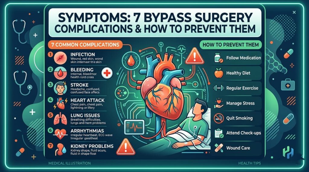
7 Bypass Surgery Complications & How to Prevent Them
Undergoing heart surgery is a major life event, and feeling anxious about the recovery process is entirely normal. Patients and their families often spend late nights researching the internet, trying to understand what to expect after leaving the operating room. While modern medicine has made this procedure incredibly safe, being aware of potential bypass surgery complications is the smartest way to ensure a smooth recovery. Knowledge is your best defense. When you know what to look out for, you can act quickly and work alongside your medical team to stay healthy. That is why top cardiothoracic experts, like Dr. Ved Prakash, emphasize patient education. By understanding your body, you can take proactive steps to protect your heart. What Are Bypass Surgery Complications? Coronary Artery Bypass Grafting (CABG) is a highly effective procedure used to treat severe heart disease. Surgeons take a healthy blood vessel from another part of your body and use it to create a detour—or bypass—around a blocked artery. This restores healthy blood flow to your heart muscle. However, because this is a major, invasive surgery, the body experiences significant stress. Bypass surgery complications refer to the medical issues, side effects, or setbacks that can occur during or after this procedure. Some complications are minor and easily treated, while others require immediate medical intervention. Let’s break down the most common ones. The 7 Major Bypass Surgery Complications 1. Bleeding and Blood Loss It is very common to experience some bleeding after any major surgical operation. However, excessive bleeding from the chest tubes or the incision site is one of the most immediate bypass surgery complications doctors watch for. To prevent severe blood loss, your surgical team will monitor you around the clock in the Intensive Care Unit (ICU) immediately following your operation. How to prevent it: Stop taking blood-thinning medications (like aspirin or warfarin) exactly when your doctor tells you to before surgery. Under the care of specialists like Dr. Ved Prakash, strict pre-surgery blood testing is conducted to minimize these risks. 2. Heart Arrhythmias (Irregular Heartbeats) After the heart is handled during surgery, it can become irritated. This irritation often leads to atrial fibrillation, a type of irregular heartbeat. This is among the most frequent bypass surgery complications, occurring in up to 30% of patients. While it can feel scary and cause a fluttering sensation in your chest, it is usually temporary and highly treatable. How to prevent it: Take all prescribed beta-blockers or anti-arrhythmic medications on schedule. Limit caffeine and stress during your initial recovery weeks at home. 3. Infections at the Incision Site Your surgeon will make an incision in your chest, and possibly in your leg or arm if a blood vessel was harvested from there. Any break in the skin carries a risk of bacterial infection. If the area becomes red, warm to the touch, or starts oozing fluid, you must act fast. Deep sternal wound infections are rare but serious bypass surgery complications that require prompt antibiotic treatment. How to prevent it: Keep your incision sites clean and dry exactly as instructed by your nursing team. Wash your hands thoroughly before touching the wound area. Monitor for fever and report any temperature spikes to your doctor immediately. 4. Kidney Function Issues During surgery, your body’s blood flow is temporarily altered, sometimes using a heart-lung bypass machine. This sudden change in blood pressure can put stress on your kidneys, leading to temporary kidney impairment. For most patients, kidney function returns to normal within a few days. However, patients with pre-existing diabetes or kidney issues face a higher risk. How to prevent it: Stay perfectly hydrated before and after your surgery according to your doctor’s guidelines. Dr. Ved Prakash and his medical team carefully monitor fluid levels and blood pressure during surgery to protect your vital organs. 5. Cognitive Decline (Brain Fog) Many patients complain of feeling confused, having memory lapses, or struggling to concentrate in the weeks following their operation. Often referred to as “pumphead,” this is one of the more frustrating bypass surgery complications. Fortunately, this cognitive decline is usually temporary. Most patients regain their full mental sharpness within six to twelve months. How to prevent it: Engage in light mental exercises like reading or solving simple puzzles during recovery. Ensure you get adequate, uninterrupted sleep every night to allow your brain to heal. 6. Blood Clots and Stroke Risk Whenever blood flow is altered, there is a risk of blood clots forming in the body. If a clot breaks loose and travels to the brain, it can cause a stroke. This is a severe risk that medical teams take extreme precautions to prevent. The risk is highest in the first few days after surgery, which is why movement and medication are heavily emphasized in the hospital. How to prevent it: Wear compression stockings provided by the hospital to keep blood flowing in your legs. Start walking as soon as your medical team gives you the green light—movement is your best defense against blood clots! 7. Pneumonia or Breathing Difficulties General anesthesia and the use of a breathing tube during surgery can leave your lungs vulnerable to fluid buildup. If this fluid is not cleared, it can lead to pneumonia—one of the most common respiratory bypass surgery complications. You will likely feel chest pain when coughing or taking deep breaths, which makes patients hesitant to breathe deeply, worsening the problem. How to prevent it: Use your spirometer (the breathing device given to you in the hospital) exactly as directed. Hold a pillow firmly against your chest when you cough; this reduces pain and protects your sternum while clearing your lungs. General Tips to Avoid Bypass Surgery Complications Minimizing the risk of bypass surgery complications doesn’t end when you leave the hospital. Your home routine is just as crucial. A successful recovery requires strict discipline, emotional support, and following your doctor’s advice to the letter. Here are the golden rules for a complication-free recovery: Follow your medication schedule strictly: Never skip doses