
Aortic Surgery

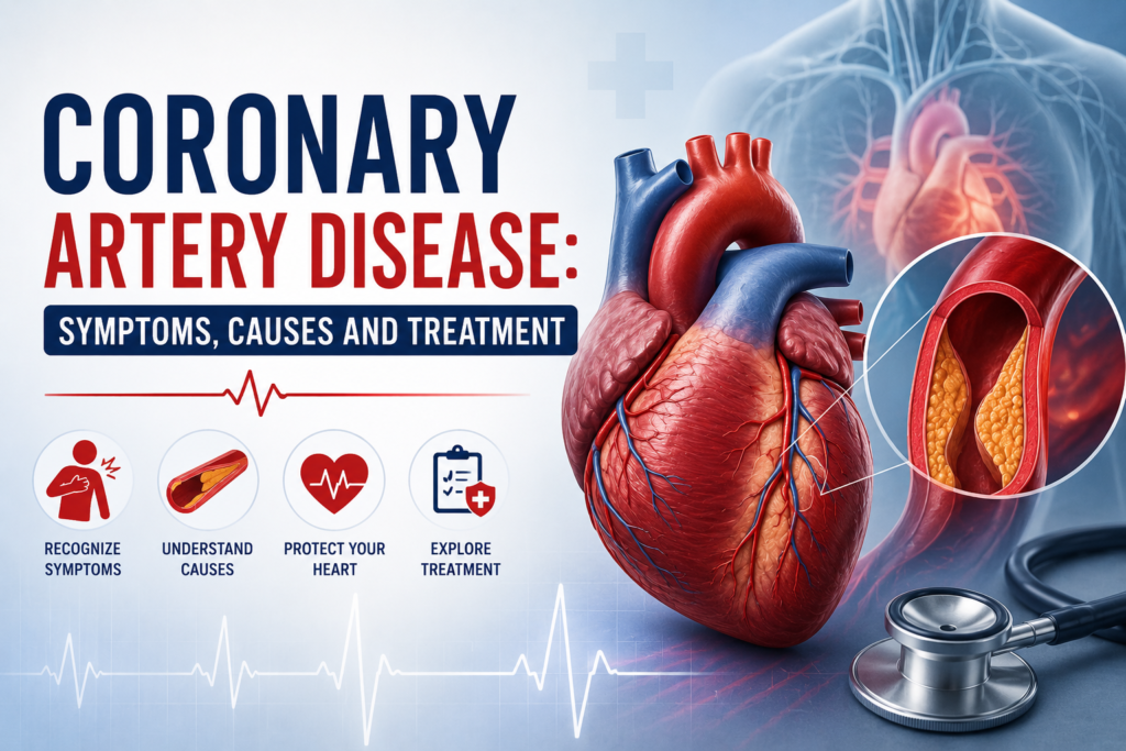
Coronary artery disease: symptoms, causes and treatment
Aortic Surgery, Cardiac Surgery

Coronary artery disease: symptoms, causes and treatment

Coronary Artery Disease (CAD) is one of the most common forms of heart disease and a leading cause of serious health complications worldwide. It develops when the coronary arteries, which supply oxygen-rich blood to the heart muscle, become narrowed or blocked due to the buildup of fatty deposits called plaque. If left untreated, Coronary Artery Disease can increase the risk of chest pain, heart attacks, heart failure, and other life-threatening conditions. The good news is that early diagnosis and proper treatment can help manage the disease effectively. Understanding the symptoms, causes, and available treatment options allows patients to take timely action and protect their long-term heart health. With expert care from an experienced cardiac surgeon like Dr. Ved Prakash, many patients can lead healthy and active lives after treatment. What Is Coronary Artery Disease? Coronary Artery Disease occurs when plaque made of cholesterol, fat, calcium, and other substances builds up inside the coronary arteries. This process, known as atherosclerosis, gradually reduces blood flow to the heart muscle. As the arteries become narrower, the heart receives less oxygen, especially during physical activity or emotional stress. Over time, severe blockages can lead to chest pain, irregular heart rhythms, or even a heart attack. In some advanced cases, cardiac surgery may be required to restore normal blood flow. Why Coronary Artery Disease Is a Serious Condition The heart works continuously to pump blood throughout the body. When blood supply is reduced because of blocked coronary arteries, the heart muscle becomes weaker and less efficient. Without timely treatment, Coronary Artery Disease may lead to serious complications such as heart attack, heart failure, or sudden cardiac arrest. Regular health check-ups and maintaining good heart health can significantly reduce these risks. Common Symptoms of Coronary Artery Disease Many people do not experience symptoms during the early stages of the disease. As the blockage increases, symptoms usually become more noticeable. The most common symptoms include: Chest pain or pressure (angina) Shortness of breath Fatigue during physical activity Pain spreading to the shoulder, arm, neck, jaw, or back Irregular heartbeat Excessive sweating Dizziness or lightheadedness Some patients, especially those with diabetes, may experience very mild or unusual symptoms. That is why regular heart screenings are important for early diagnosis. Warning Signs of a Heart Attack A heart attack can occur when a coronary artery becomes completely blocked. Recognizing the warning signs can save lives. Seek immediate medical attention if you experience: Severe chest pain lasting more than a few minutes Pain radiating to the left arm, jaw, or back Sudden shortness of breath Cold sweating Nausea or vomiting Sudden weakness or fainting Prompt treatment greatly improves the chances of recovery and reduces damage to the heart muscle. What Causes Coronary Artery Disease? Several factors contribute to the development of Coronary Artery Disease. While some risk factors cannot be changed, many can be controlled through healthy lifestyle choices. The most common causes include: High cholesterol High blood pressure Smoking Diabetes Obesity Physical inactivity Unhealthy diet Excessive alcohol consumption Chronic stress Family history of heart disease Increasing age Making positive lifestyle changes can slow disease progression and improve overall heart health. Who Is at Higher Risk? Certain individuals have a greater chance of developing Coronary Artery Disease. Understanding your personal risk helps you take preventive measures before symptoms appear. People at higher risk include older adults, smokers, individuals with diabetes, patients with uncontrolled high blood pressure or cholesterol, overweight individuals, and those with a family history of heart disease. During routine consultations, Dr. Ved Prakash evaluates these risk factors carefully and recommends personalized strategies to lower the chances of future heart complications through lifestyle modifications, medications, or advanced cardiac surgery when necessary. How Is Coronary Artery Disease Diagnosed? Early diagnosis is essential for preventing complications and protecting heart health. If your doctor suspects Coronary Artery Disease, several tests may be recommended to evaluate blood flow, heart function, and the severity of arterial blockages. The choice of tests depends on your symptoms, medical history, age, and overall health. A detailed evaluation helps the cardiac surgeon or cardiologist recommend the most appropriate treatment plan. Some commonly used diagnostic tests include: Electrocardiogram (ECG) Echocardiogram (Echo) Exercise stress test CT Coronary Angiography Coronary Angiography Blood tests for cholesterol and blood sugar Cardiac MRI in selected cases These investigations help identify narrowed arteries and determine whether medications, minimally invasive procedures, or cardiac surgery are required. Treatment Options for Coronary Artery Disease The treatment of Coronary Artery Disease depends on the severity of the blockage, the patient’s symptoms, and overall heart health. In many cases, a combination of lifestyle changes, medications, and medical procedures provides the best results. The primary goals of treatment are to improve blood flow, relieve symptoms, prevent heart attacks, and enhance quality of life. Lifestyle Changes That Support Heart Health Healthy lifestyle habits are the foundation of managing Coronary Artery Disease. Even patients who require medications or surgery benefit greatly from long-term lifestyle improvements. Doctors commonly recommend: Eating a balanced heart-healthy diet Exercising regularly under medical guidance Maintaining a healthy body weight Quitting smoking completely Limiting alcohol consumption Controlling stress through yoga or meditation Getting adequate sleep Managing diabetes, blood pressure, and cholesterol These simple habits can slow disease progression and significantly improve heart health over time. Medications Used for Coronary Artery Disease Many patients can successfully manage their condition with medications, especially during the early stages of the disease. Depending on individual needs, doctors may prescribe: Blood-thinning medications Cholesterol-lowering medicines (statins) Blood pressure medications Medicines to reduce chest pain (anti-anginal drugs) Beta-blockers ACE inhibitors or ARBs Patients should always take medications exactly as prescribed and avoid stopping them without consulting their healthcare provider. When Is Angioplasty Recommended? For patients with localized coronary artery blockages, angioplasty may be an effective treatment option. During this minimally invasive procedure, a small balloon is used to widen the narrowed artery, and a stent is often placed to keep the artery open. Angioplasty usually offers quicker recovery than surgery and is suitable for selected patients

Aortic Surgery

What Is an Aortic Aneurysm? Silent, Dangerous — and Treatable

An aortic aneurysm is a ballooning in the wall of the aorta — the body’s main artery — and the most dangerous thing about it is that it causes no symptoms until it is on the verge of rupturing. Rupture of an aortic aneurysm is one of the most lethal events in medicine, with mortality exceeding 80% even with emergency surgery. The goal — always — is to find it and treat it before that moment. Dr. Ved Prakash, Director of CTVS at Yatharth Super Speciality Hospitals, Greater Noida, explains what causes an aortic aneurysm, when it needs surgery, and what the surgical options are. What Is an Aortic Aneurysm? The aorta is the largest artery in the body — rising from the heart, arching through the chest, and running down through the abdomen to supply the entire lower body. The normal aortic diameter is approximately 2.0–3.0 cm. An aortic aneurysm is an abnormal widening of the aorta to more than 1.5 times its normal diameter — defined as greater than 3.0 cm in the abdominal aorta or greater than 4.5 cm in the thoracic aorta. As an aortic aneurysm enlarges, the aortic wall becomes progressively thinner and weaker. At a critical size, the wall can rupture — and the outcome is catastrophic. Types of Aortic Aneurysm Abdominal Aortic Aneurysm (AAA) The most common type — a ballooning in the portion of the aorta that runs through the abdomen. AAAs occur primarily in men over 65 with a history of smoking, high blood pressure, and high cholesterol. They are almost always discovered incidentally on an ultrasound or CT scan ordered for something else entirely — because they rarely cause symptoms. Thoracic Aortic Aneurysm (TAA) Aortic aneurysm in the chest — affecting the ascending aorta (rising from the heart), the arch, or the descending aorta. TAAs are more commonly associated with genetic conditions including Marfan syndrome and bicuspid aortic valve, and can affect patients at younger ages than AAAs. What Causes an Aortic Aneurysm? Atherosclerosis — the primary cause of AAA. Years of high blood pressure, high cholesterol, and smoking progressively weaken the aortic wall. Hypertension — the most important modifiable risk factor. Controlling blood pressure directly slows aneurysm growth. Smoking — doubles aortic aneurysm risk and accelerates growth rate significantly. The single most effective prevention is smoking cessation. Marfan syndrome and related connective tissue disorders — inherently weak aortic wall tissue; aneurysms can develop at younger ages and at smaller sizes than in the general population. Bicuspid aortic valve — associated with progressive ascending aortic enlargement; regular echocardiographic surveillance of the aortic diameter is essential. Family history — first-degree relatives of AAA patients have a 10–15% lifetime risk; screening ultrasound is recommended. Aortic Aneurysm Symptoms — Why It Is Called a Silent Killer The vast majority of aortic aneurysms produce no symptoms until they are large or rupturing. When symptoms do occur: Deep, constant back or abdominal pain — often mistaken for musculoskeletal pain. In the context of a known aortic aneurysm, this is a warning sign of rapid expansion or contained leak — requiring emergency assessment. Pulsating abdominal mass — a pulsating sensation in the centre of the abdomen, felt by the patient or detected on physical examination. Most clearly felt in thin patients. Hoarseness or difficulty swallowing — from a thoracic aneurysm pressing on the recurrent laryngeal nerve or oesophagus. Sudden, severe tearing abdominal or back pain with collapse is rupture until proven otherwise — call emergency services immediately. This is different from the presentation of aortic dissection, which classically causes chest pain — though both are surgical emergencies. When Does an Aortic Aneurysm Need Surgery? The decision is based primarily on size — larger aneurysms have exponentially higher annual rupture risk: AAA Diameter Annual Rupture Risk Recommendation 3.0–4.4 cm <0.5% Ultrasound surveillance every 12 months 4.5–5.4 cm 1–5% Surveillance every 6 months + surgical review 5.5 cm and above 10–25% Surgery recommended Growing >1 cm per year (any size) High Surgery recommended regardless of size EVAR vs Open Surgery — What Are the Options? EVAR — Endovascular Aneurysm Repair A stent-graft is delivered through the femoral arteries in the groin and positioned inside the aneurysm, lining it and excluding it from the circulation. No abdominal incision. Hospital stay 2–3 days. Recovery 2–3 weeks. Requires annual CT surveillance to check the stent-graft remains in position and there is no re-pressurisation (endoleak). Suitable for most infrarenal AAAs with appropriate anatomy. Open Surgical Repair The aneurysm is exposed through an abdominal incision, clamped, and replaced with a synthetic Dacron graft. More invasive — hospital stay 7–10 days, recovery 6 weeks — but provides a definitive, lifelong repair that does not require ongoing CT surveillance. Required when anatomy is not suitable for EVAR, or for juxtarenal/suprarenal aneurysms. TEVAR — Thoracic Endovascular Aortic Repair The endovascular equivalent of EVAR for descending thoracic aortic aneurysms — a stent-graft relined the diseased thoracic aorta through femoral artery access. For patients in Delhi NCR with a newly diagnosed or enlarging aortic aneurysm, aortic surgery in Delhi NCR at Yatharth Hospital covers the full range — EVAR, TEVAR, and open surgical repair. CT reports can be shared via WhatsApp for a pre-assessment before your visit. Frequently Asked Questions — What Is Aortic Aneurysm What is an aortic aneurysm and is it always fatal? An aortic aneurysm is an abnormal ballooning of the aortic wall. It is not immediately fatal — most detected aneurysms are small and slow-growing and can be treated safely with elective surgery before rupture. A ruptured aneurysm carries mortality above 80%. Finding it early is what saves lives. At what size does an aortic aneurysm need surgery? Surgery is generally recommended when an abdominal aortic aneurysm reaches 5.5 cm or when it grows more than 1 cm in 12 months. Below this, surveillance ultrasound every 6–12 months is the standard approach. What is EVAR and how is it different from open surgery? EVAR delivers a stent-graft through the groin without opening the

Aortic Surgery

Aortic Dissection — Symptoms, Emergency Surgery and What Families Need to Know

If someone near you has sudden, severe tearing chest or back pain — call emergency services immediately and do not drive. Aortic dissection is one of the few conditions where minutes determine survival. Aortic dissection is a tear in the inner wall of the aorta that allows blood to surge between the layers of the aortic wall — and it is the most time-critical emergency in cardiac surgery. Unlike most cardiac conditions, aortic dissection symptoms have a characteristic that distinguishes them from almost everything else: the pain is at its absolute worst the moment it begins. Dr. Ved Prakash, Director of CTVS at Yatharth Super Speciality Hospitals, Greater Noida, explains aortic dissection symptoms, the classification that determines treatment urgency, and what family members need to know when every minute counts. What Is Aortic Dissection? The aortic wall has three layers. In aortic dissection, the innermost layer (intima) develops a tear — and blood under arterial pressure forces through that tear and strips the layers apart along the length of the aorta, creating a false channel (false lumen) alongside the true blood channel. As this false channel extends, it can compress the true channel — cutting off blood supply to organs. It can propagate backwards toward the heart — causing severe aortic regurgitation or cardiac tamponade. It can block the openings of coronary arteries — causing a simultaneous heart attack. And it can rupture through the outer wall — causing catastrophic internal haemorrhage. This cascade can progress from tear to death in under an hour in the worst cases. Aortic Dissection Symptoms — The Defining Characteristics Tearing or Ripping Chest or Back Pain — Maximum at Onset This is the single most important aortic dissection symptom, and what separates it clinically from almost every other cardiac emergency. Patients describe it as tearing, ripping, or like something exploding inside the chest. It is at maximum intensity from the very first second — not building gradually. Most patients describe it as the worst pain they have ever experienced in their life. It often radiates to the back, between the shoulder blades. Migrating Pain As the dissection extends down the aorta, the pain follows it — moving from the chest to the back, then toward the abdomen. Migrating pain is highly specific for aortic dissection and should always trigger immediate emergency assessment with CT angiography. Blood Pressure Difference Between the Two Arms A systolic blood pressure difference of more than 20 mmHg between the right and left arm — caused by the dissection narrowing or blocking the artery to one arm — is a clinical sign strongly associated with aortic dissection. In emergency settings, blood pressure should always be measured in both arms when dissection is suspected. Neurological Symptoms Sudden weakness, numbness, or paralysis of an arm or leg — caused by the dissection compromising blood supply to the spinal cord or carotid arteries. When aortic dissection presents with stroke symptoms, it is particularly dangerous because thrombolytic drugs (clot-busters given in stroke) are absolutely contraindicated — they would cause fatal haemorrhage in a dissection. Absent Pulse in One Limb If the dissection extends into a major branch artery, the pulse may disappear in one arm or leg. Stanford Classification — Type A vs Type B The most important classification in aortic dissection. Everything — urgency, treatment, mortality — hinges on which type it is. Type A — Surgical Emergency Any dissection involving the ascending aorta (the section of aorta rising from the heart) — regardless of where the original tear is. Mortality is approximately 1–2% per hour without surgery. Emergency surgical replacement of the ascending aorta must be performed as soon as the operating team can be assembled — ideally within 2 hours of diagnosis. This is one of the most technically demanding emergency operations in cardiac surgery. Type B — Medical Management Initially Dissection involving only the descending aorta, below the left subclavian artery, not extending into the ascending aorta. Uncomplicated Type B is managed medically — strict blood pressure and heart rate control in a cardiac ICU with IV medications. Complicated Type B — with evidence of organ malperfusion, rapidly expanding false lumen, or threatened rupture — requires urgent endovascular repair (TEVAR). What to Tell the Emergency Room Team If you suspect aortic dissection in yourself or a family member, say these specific words to the emergency team: “Sudden, severe chest or back pain that was worst immediately when it started” “The pain feels like tearing or ripping” “They have high blood pressure” (or Marfan syndrome, or a known aortic aneurysm, if applicable) This prompts immediate CT aortic angiography — the definitive test that confirms or excludes aortic dissection within minutes. Do not let the team assume heart attack and give thrombolytics without a CT scan — in aortic dissection, this would be fatal. Risk Factors for Aortic Dissection Hypertension — the most common risk factor. Poorly controlled blood pressure progressively weakens the aortic wall. Marfan syndrome — inherently weak aortic wall; dissection can occur at younger ages and smaller aortic diameters. Bicuspid aortic valve — associated with ascending aortic enlargement and dissection risk. Existing aortic aneurysm — an enlarged aorta is at higher risk of dissection; learn more about aortic aneurysm. Pregnancy — third trimester and peripartum period; rare but important cause of Type A dissection in young women. Emergency Surgery for Type A Aortic Dissection The operation is performed under general anaesthesia on cardiopulmonary bypass. The ascending aorta is replaced with a synthetic Dacron graft. If the dissection extends into the aortic arch, deep hypothermic circulatory arrest — cooling the body to 18°C and temporarily stopping circulation — allows the surgeon to repair the arch safely. If the aortic valve root is involved, it may be repaired or replaced simultaneously. Total operating time: 4–8 hours. This is among the highest-stakes operations in all of surgery — and one that Dr. Ved Prakash performs as part of the aortic surgery programme at Yatharth Hospital. Frequently Asked Questions — Aortic Dissection

Scroll to Top