
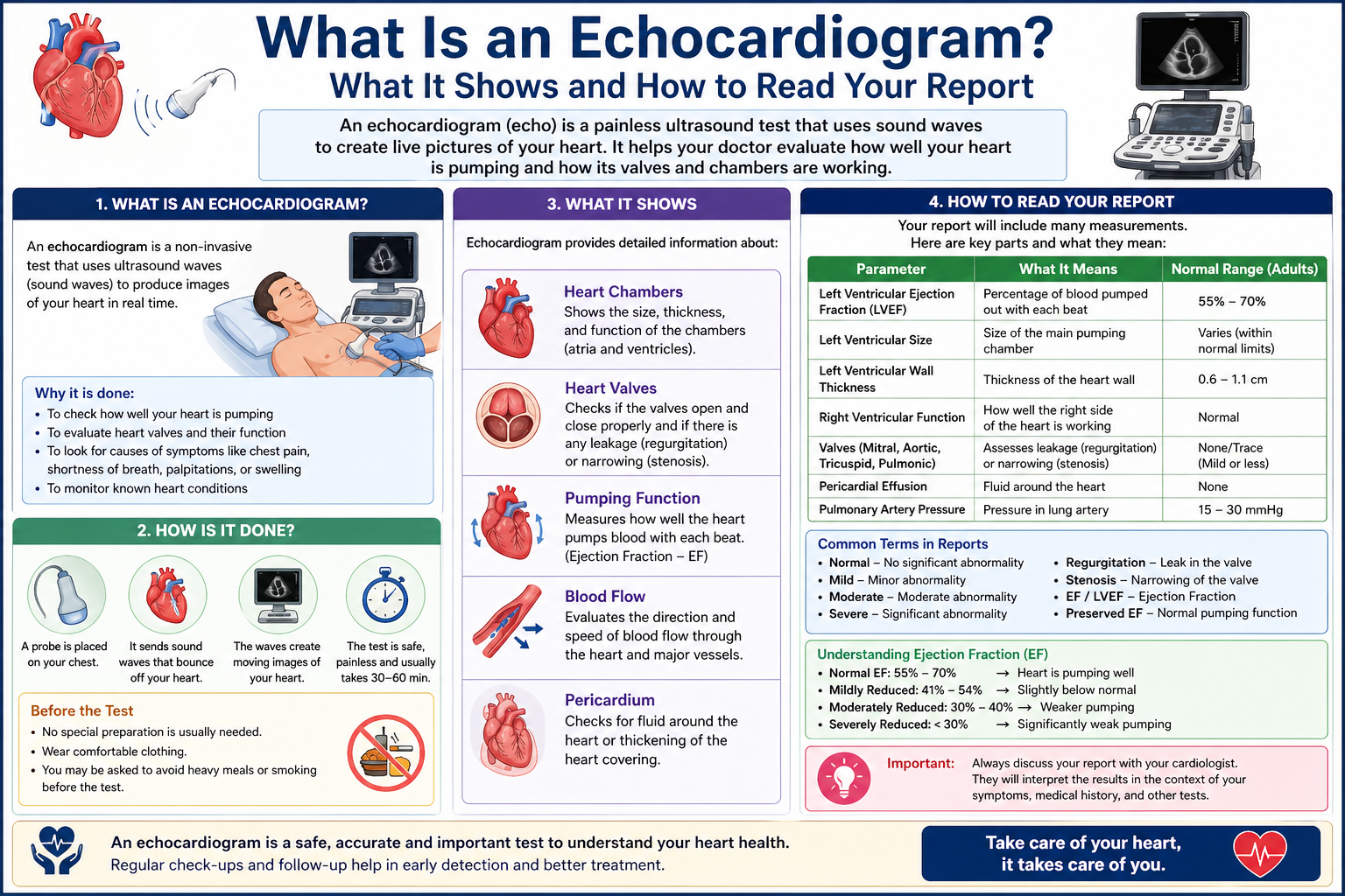
 What Is an Echocardiogram? Echocardiogram is an ultrasound scan of your heart — the single most important investigation in cardiac medicine, and the test Dr. Ved Prakash reviews before making any surgical recommendation. If you have been asked to get one, or if you already have a report in hand and cannot understand it, this guide explains what an echocardiogram shows, how to read the key numbers, and what different findings mean for your treatment.
Dr. Ved Prakash is Director of Cardiothoracic and Vascular Surgery at Yatharth Super Speciality Hospitals, Greater Noida, and has been reviewing echocardiograms as part of surgical planning for over 8 years across Medanta, Narayana, and Sarvodaya.
What Is an Echocardiogram — In Plain Language
An echocardiogram uses sound waves at a frequency too high for human hearing — ultrasound — to produce real-time, moving images of your heart. The probe is placed on your chest and the reflected sound waves are converted into pictures on a screen. You see the heart beating, the walls contracting, the valves opening and closing — all in motion.
Unlike an ECG, which only shows electrical activity, an echocardiogram shows structure. It answers the questions an ECG cannot: Is the heart pumping effectively? Are the valves leaking or narrowed? Are any walls of the heart not moving properly? Is there fluid around the heart?
A standard echocardiogram takes 20–30 minutes, uses no radiation, and requires no preparation. It is painless.
What Does an Echocardiogram Show?
A single echocardiogram provides more clinical information about the heart than any other non-invasive test:
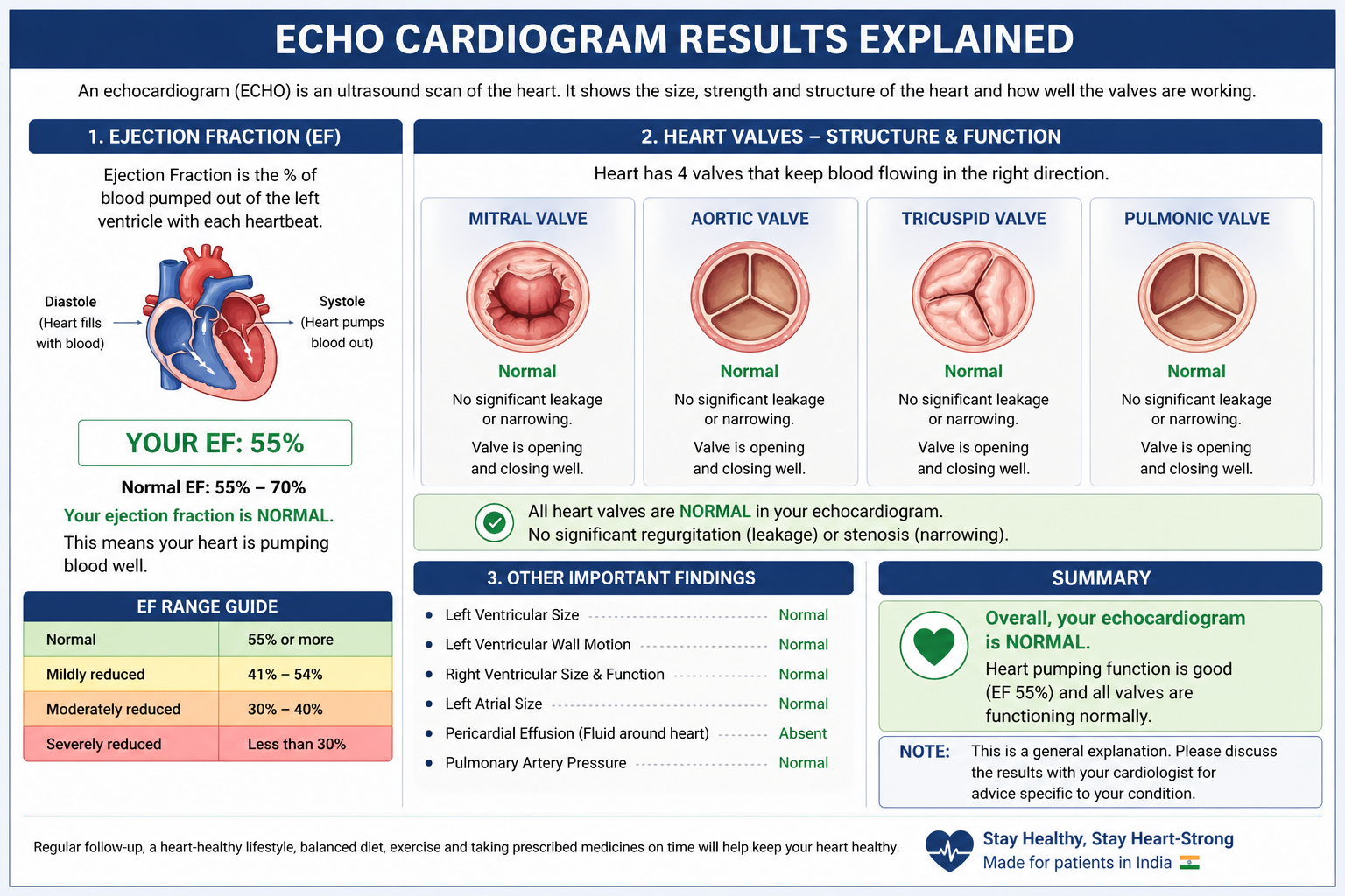
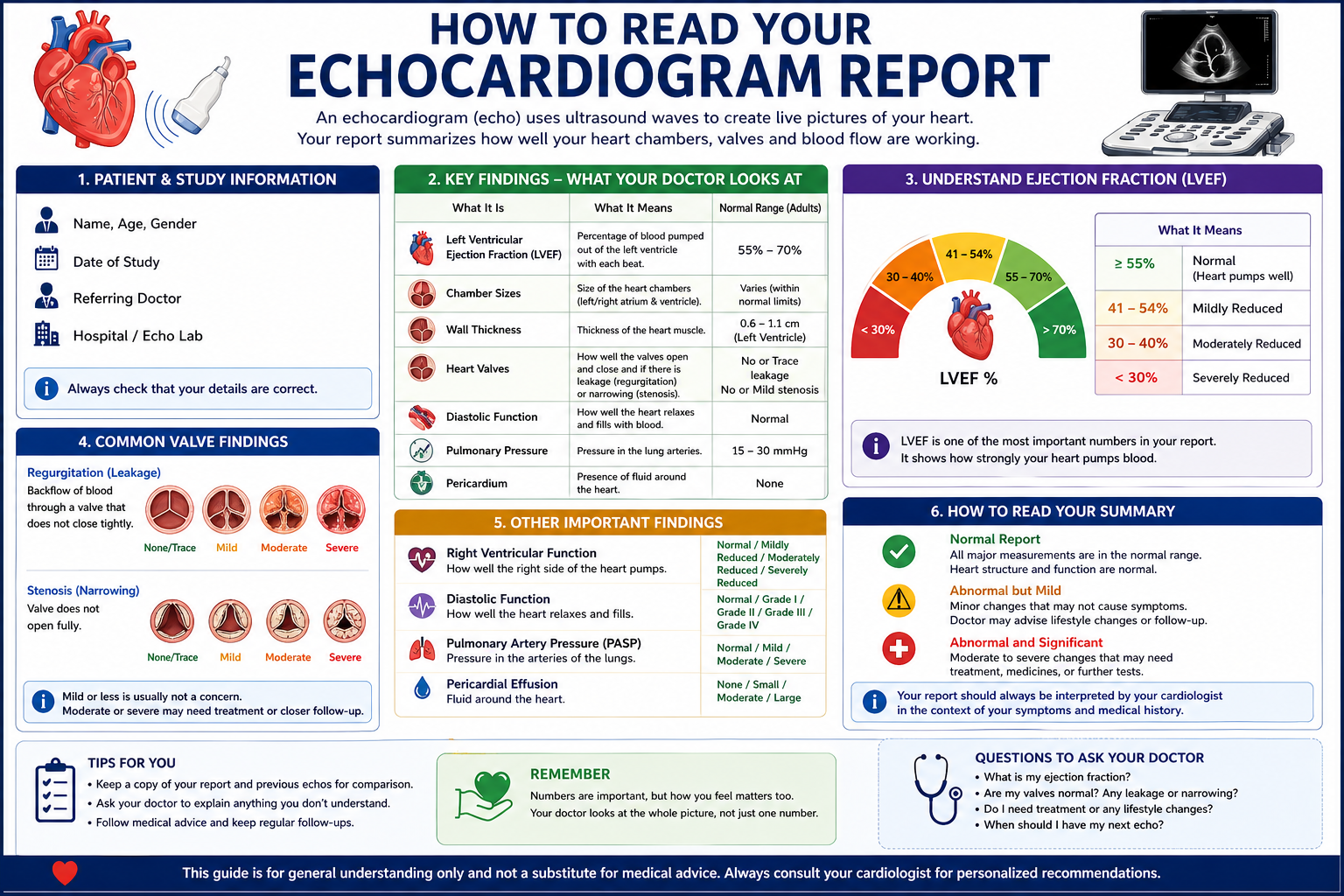
- Ejection fraction (EF) — the pumping power of the left ventricle. The most important number on the report.
- Wall motion — whether each segment of the heart wall is contracting normally. Areas that are not moving indicate past heart attack damage or ongoing ischaemia.
- Valve function — whether each of the four valves is opening fully (stenosis) or closing completely (regurgitation), and how severe the problem is.
- Chamber size — whether the heart chambers have enlarged from chronic pressure or volume overload.
- Pericardial effusion — fluid around the heart that can compress cardiac function if significant.
- Congenital defects — holes between chambers (ASD, VSD) or structural abnormalities present from birth.
How to Read Your Echocardiogram Report
Ejection Fraction (EF)
This is the most watched number on any echocardiogram report. It measures what percentage of blood in the left ventricle is pumped out with each heartbeat.
- 55–70%: Normal. The heart is pumping well.
- 40–54%: Mildly reduced. Medication review and repeat echocardiogram in 3–6 months is standard.
- 30–39%: Moderately reduced. Specialist review and treatment required.
- Below 30%: Severely reduced. Significant heart failure. This number changes what surgery is recommended, when it is done, and how
Valve Severity Grading
Every abnormal valve on the echocardiogram is graded as mild, moderate, or severe. Mild disease is monitored — repeat echo in 1–2 years. Moderate disease means more frequent follow-up — every 6–12 months. Severe disease with symptoms, or with evidence of chamber enlargement on the same echocardiogram, is a surgical discussion. This is the threshold at which a consultation with a cardiac surgeon — specifically about heart valve surgery — becomes necessary.
Wall Motion Abnormality
Each segment of the left ventricular wall is scored: 1 = normal movement, 2 = reduced movement (hypokinesia), 3 = no movement (akinesia), 4 = bulging outward (dyskinesia — indicates a ventricular aneurysm). Any score above 1 in multiple segments means past heart attack damage or active ischaemia from a blocked artery.
 Types of Echocardiogram
Transthoracic Echocardiogram (TTE)
The standard test. Probe on the chest wall. Completely painless. No preparation. Done in 20–30 minutes. This is what most patients have when their cardiologist orders an “echo”.
Transoesophageal Echocardiogram (TOE)
A probe is passed into the food pipe under sedation — giving far clearer images of the mitral valve, the back of the heart, and the left atrium. Used before mitral valve surgery, in suspected endocarditis, and to look for clots in the left atrial appendage before cardioversion.
Stress Echocardiogram
An echocardiogram is taken at rest, then immediately after exercise on a treadmill. Wall motion abnormalities that only appear during exertion reveal blocked arteries that are not obvious on a resting echo — useful when symptoms suggest angina but the resting study is normal.
When Does an Echocardiogram Lead to Surgery?
Not every abnormal echocardiogram leads to surgery. But the following findings typically trigger a surgical consultation:
- Ejection fraction below 35% with symptoms of breathlessness or fatigue
- Severe aortic stenosis — valve area below 1.0 cm² with any symptoms
- Severe mitral regurgitation with increasing left ventricular dimensions
- Significant ASD or VSD with right heart enlargement on the same study
- Multiple wall motion abnormalities suggesting active ischaemia from blocked coronary arteries
If your echocardiogram has raised a concern and you want a surgical opinion, you can share your echo report online for a consultation with Dr. Ved Prakash — without having to travel first.
Frequently Asked Questions — What Is an Echocardiogram
What is an echocardiogram and is it the same as an ECG?
No — they are completely different. An ECG records electrical signals. An echocardiogram uses ultrasound to show the heart’s structure and movement — valves, walls, chambers, fluid. An echocardiogram gives far more diagnostic information than an ECG.
What does ejection fraction mean on an echocardiogram?
Ejection fraction is the percentage of blood pumped out by the left ventricle per beat. Normal is 55–70%. Below 40% indicates significant heart failure and directly influences decisions about surgery type and timing.
What does a normal echocardiogram report look like?
EF 55–70%, all four valves functioning normally, no wall motion abnormalities, normal chamber sizes, no pericardial effusion. If your report shows all of these, your heart structure and function are normal.
Is an echocardiogram safe?
Completely safe. Ultrasound only — no radiation. Painless and preparation-free. Safe for newborns, pregnant women, and the elderly without any risk.
Dr. Ved Prakash | Director, CTVS — Yatharth Super Speciality Hospitals, Greater Noida
📞 +91-9355255106 |
📧 drvedprakash@gmail.com |
Book Appointment →