
 Coronary angiography is the definitive test for identifying blockages in the heart arteries — the investigation that gives every cardiologist and cardiac surgeon the exact road map needed to plan the right treatment. If you have been referred for coronary angiography, or if you already have a report and cannot make sense of it, Dr. Ved Prakash, Director of CTVS at Yatharth Super Speciality Hospitals, Greater Noida, explains what the procedure involves, what the results mean, and what comes next.
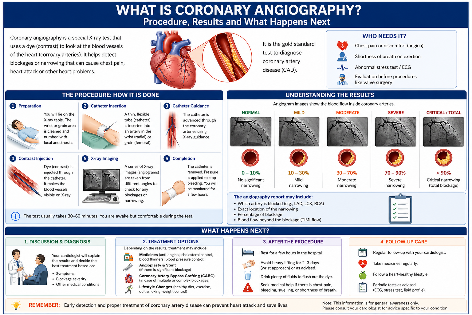
What Is Coronary Angiography?
Coronary angiography is a catheter-based procedure in which a special dye (contrast agent) is injected directly into the coronary arteries while X-ray images are captured continuously. The dye makes the inside of the arteries visible — showing exactly where blockages are, how severe they are, and how many arteries are affected.
It is performed in a cardiac catheterisation laboratory (cath lab), takes 30–45 minutes, and is done under local anaesthesia. You remain awake throughout. Most hospitals now use the radial artery in the wrist for access — which means less discomfort and same-day or next-morning discharge in most cases.
Why Has Your Doctor Recommended Coronary Angiography?
Coronary angiography is recommended when:
- Chest pain on exertion suggests blocked coronary arteries (angina)
- An ECG, stress test, or echocardiogram has shown changes that need further investigation
- You are being assessed before major heart surgery — such as valve replacement — and need coronary status confirmed
- You have had a heart attack and require urgent identification of the blocked artery
- A CT coronary angiogram has found blockages that need catheter-based confirmation before treatment
What Happens During Coronary Angiography — Step by Step
Preparation
You fast for 4–6 hours before the procedure. An IV line is placed in your arm. The access site — usually your wrist — is cleaned and numbed with local anaesthetic.
Catheter Insertion
A thin, flexible catheter is inserted into the radial artery and guided up through the arm and chest to the opening of each coronary artery. This is done under live X-ray guidance. You feel pressure but no pain.
Dye Injection and Imaging
A small amount of contrast dye is injected into each coronary artery. For 10–15 seconds you will feel a warm flush through the chest — this is completely normal and passes quickly. X-ray images are captured from multiple angles as the dye flows through the arteries.
Catheter Removal and Recovery
The catheter is removed. A compression band is placed over the wrist access site. Within an hour you can sit up and eat. Most patients go home the same evening or the following morning.
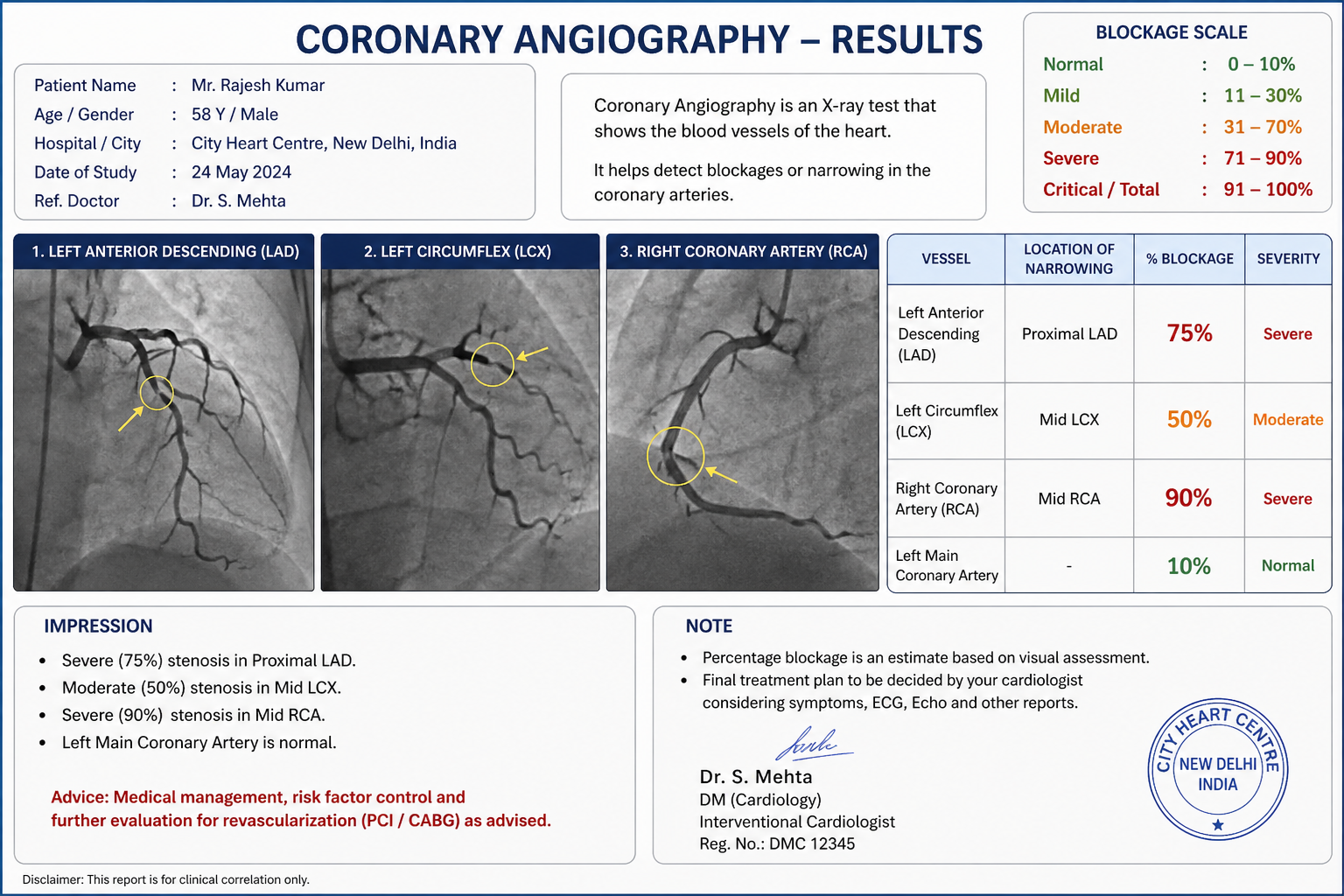
How to Read Your Coronary Angiography Report
This is where most patients feel lost — and where the most important decisions get made. Here is what the numbers mean.
Percentage Blockage (Stenosis)
| Stenosis | What It Means | Typical Action |
| 0–49% | Mild narrowing — does not significantly restrict flow | Medication and lifestyle. No procedure needed. |
| 50–69% | Moderate — may or may not restrict flow | FFR pressure wire test to confirm significance |
| 70–90% | Significant — restricts blood flow | Treatment recommended: stent or bypass |
| 90–99% | Critical — very high heart attack risk | Urgent treatment |
| 100% | Complete blockage (total occlusion) | Depends on duration and viable muscle at risk |
Which Arteries Are Named in the Report
- LAD (Left Anterior Descending): Supplies the front of the heart. The most important coronary artery — sometimes called the “widow maker” when severely blocked.
- LCx (Left Circumflex): Supplies the side and back of the left ventricle.
- RCA (Right Coronary Artery): Supplies the right ventricle and the back of the left ventricle.
- Left Main: The trunk from which the LAD and LCx both arise. A significant left main blockage is treated as a surgical situation in most cases.
What Happens After Coronary Angiography If Blockages Are Found?
Single, simple blockage: May be treated with angioplasty and stenting in the same session or shortly after — particularly if the patient is non-diabetic and the anatomy is straightforward.
Multiple blockages or complex anatomy: The angiography images are reviewed by a Heart Team — interventional cardiologist and cardiac surgeon together. They use the SYNTAX score to assess complexity and recommend either angioplasty or bypass surgery based on what gives the best long-term result for that specific anatomy.
Left main disease: A cardiac surgical review is mandatory before any decision. Bypass surgery is recommended in most left main cases.
No significant blockages: Coronary artery disease is effectively ruled out. Your symptoms need evaluation for other causes.
You Do Not Have to Decide Immediately
One thing many patients do not know: if angiography is being done electively and blockages are found, you are not obligated to accept stenting on the same table. You have every right to review the images, take the report, and seek an independent review of your angiogram from a cardiac surgeon before deciding between bypass surgery and angioplasty. This is not delay — it is good medicine.
Frequently Asked Questions — What Is Coronary Angiography
What is coronary angiography and is it dangerous?
Coronary angiography is a catheter-based diagnostic test that uses dye and X-ray to reveal coronary artery blockages. It is very safe — serious complications occur in less than 0.1% of elective procedures. Local anaesthesia, 30–45 minutes, wrist access in most cases.
What does 70% blockage on angiography mean?
The artery is narrowed to 30% of its normal diameter — significantly restricting blood flow. Most cardiologists and cardiac surgeons recommend treatment for 70%+ blockages in major coronary arteries. Whether treatment is stenting or bypass surgery depends on the number of vessels involved and the overall anatomy.
Is coronary angiography painful?
Not painful. The wrist or groin is numbed with local anaesthetic before the catheter is inserted. Patients feel a brief warm flush when the dye is injected — 10–15 seconds — which is normal. Most patients find angiography considerably less uncomfortable than they anticipated.
What happens after coronary angiography if a blockage is found?
A single simple blockage may be stented in the same session. Multiple or complex blockages are reviewed by a Heart Team who recommend angioplasty or bypass surgery based on the SYNTAX score. You are not obligated to accept treatment immediately.
Dr. Ved Prakash | Director, CTVS — Yatharth Super Speciality Hospitals, Greater Noida
📞 +91-9355255106 |
📧 drvedprakash@gmail.com |
Book Appointment →