Author name: Atul Manori

Uncategorized

Paediatric Heart Surgeon in Noida — Congenital Heart Surgery at Yatharth Hospital

If your child has been diagnosed with a congenital heart defect and you are looking for a paediatric heart surgeon in the Noida or Greater Noida area — Yatharth Super Speciality Hospitals, Sector 110, Greater Noida is 25–30 minutes from Noida City Centre. Dr. Ved Prakash, Director of CTVS, performs the full range of paediatric and congenital cardiac surgery here — from neonatal emergencies to planned repairs in infants, children, and adolescents.   Congenital Heart Conditions Treated ASD (Atrial Septal Defect) Surgical patch closure · Device closure via catheter for suitable anatomy VSD (Ventricular Septal Defect) Surgical patch closure from 3–4 months · Device closure for muscular VSDs TOF (Tetralogy of Fallot) Complete intracardiac repair · VSD patch + right ventricular outflow tract relief TGA (Transposition of Great Arteries) Arterial switch operation — neonatal surgery within first 2 weeks of life Coarctation of Aorta Surgical repair · End-to-end anastomosis or patch aortoplasty TAPVC (Total Anomalous Pulmonary Venous Connection) Emergency neonatal repair · Pulmonary vein redirection to left atrium PDA (Patent Ductus Arteriosus) Surgical ligation · Device closure Complex single-ventricle disease Bidirectional Glenn shunt · Fontan completion Why Families From Noida Choose Yatharth Hospital 25–30 minutes from Noida City Centre — no Delhi traffic, expressway route Senior CTVS surgeon on-site — Dr. Ved Prakash has performed paediatric cardiac surgery from Medanta, Narayana, Sarvodaya, and Yatharth Neonatal cardiac surgical capability — for infants requiring surgery in the first days or weeks of life Dedicated paediatric cardiac pathway — separate from adult cardiac patients No waiting list — urgent and elective paediatric cases scheduled without the 6–12 week delays seen at government centres Ayushman Bharat, CGHS, ECHS, and all insurance accepted What to Do First — Before Travelling Share your child’s echocardiogram report via WhatsApp to +91-9355255106 — Dr. Ved Prakash reviews and advises on urgency and surgical plan If a foetal echocardiogram has detected a defect before birth — share the report and plan delivery accordingly Book OPD — confirm appointment before making the trip For more on the most common conditions treated, read about VSD in babies or visit the paediatric heart surgery service page. Frequently Asked Questions Is there a paediatric heart surgeon in Noida? Yes — Dr. Ved Prakash at Yatharth Hospital, Greater Noida (25–30 min from Noida City Centre) performs the full range of paediatric and congenital heart surgery including ASD, VSD, TOF, TGA, coarctation, TAPVC, and neonatal cardiac surgery. What age can children have heart surgery at Yatharth Hospital? From the neonatal period — including critical surgeries for TGA and TAPVC in the first days of life — through infancy, childhood, and adolescence. Age and weight are not barriers when surgery is medically necessary. How do I share my child’s echo report with Dr. Ved Prakash? WhatsApp the report to +91-9355255106. A pre-assessment is provided including likely surgical plan and timing — before you travel. Dr. Ved Prakash | Director, CTVS — Yatharth Super Speciality Hospitals, Greater Noida 📞 +91-9355255106  | 📧 drvedprakash@gmail.com  | Book a Consultation →

congenital heart defect in children
Uncategorized

Congenital Heart Defect in Children — A Parent’s Complete Guide

A congenital heart defect in children is a structural problem with the heart that is present from birth — and it is more common than most parents realise. Approximately 9 in every 1,000 babies born in India are affected. If your child has just been diagnosed, the most important thing to understand first is this: the majority of congenital heart defects can be completely corrected with surgery. Dr. Ved Prakash, Director of CTVS at Yatharth Super Speciality Hospitals, Greater Noida, has been performing paediatric heart surgery since 2017 — operating on newborns, infants, and children with a wide range of congenital conditions. This guide is written for parents — to give you clear, honest answers in the most stressful weeks of your family’s life.   What Is a Congenital Heart Defect? A congenital heart defect is any abnormality in the heart’s structure that develops during pregnancy. The heart forms in the first 8 weeks of fetal development — and during this complex process, variations can occur in the walls between chambers, the heart valves, or the major blood vessels leaving the heart. Some defects are simple — a small hole between chambers that may close on its own. Others are complex — involving multiple structural abnormalities that require surgical correction in the first days or weeks of life. What Causes a Congenital Heart Defect? In most cases, no single identifiable cause is found. Known risk factors include: Chromosomal conditions such as Down’s syndrome (trisomy 21) — associated with ASD, VSD, and AV canal defects Rubella (German measles) infection in the mother during the first trimester Maternal diabetes — poorly controlled blood sugar during pregnancy increases risk Certain medications taken during pregnancy A family history of congenital heart disease — though most cases occur without family history Parents should not blame themselves. Congenital heart defects are not caused by anything the mother ate, did, or felt during pregnancy in the vast majority of cases. The Most Common Congenital Heart Defects in Children ASD — Atrial Septal Defect A hole in the wall (septum) between the two upper chambers (atria) of the heart. Small ASDs often close on their own in the first few years of life. Larger ASDs allow too much blood to flow from the left side to the right side — over time causing the right side of the heart to enlarge and leading to pulmonary hypertension if untreated. Larger ASDs are closed surgically or with a catheter-based device — both are extremely safe and effective. VSD — Ventricular Septal Defect The most common congenital heart defect — a hole between the two lower chambers (ventricles). Small VSDs often close spontaneously in infancy. Large VSDs cause significant left-to-right shunting of blood, leading to poor weight gain, breathlessness with feeding, frequent chest infections, and eventually pulmonary hypertension. Surgical closure restores completely normal circulation. TOF — Tetralogy of Fallot A combination of four structural defects — a large VSD, obstruction to blood flow from the right ventricle to the lungs, an overriding aorta, and right ventricular thickening. The combination means oxygen-depleted (blue) blood is pumped to the body — causing cyanosis (bluish discolouration of the lips and fingernails). This is the classic “blue baby” condition. Complete surgical repair is typically performed between 3 and 6 months of age with excellent long-term outcomes. TAPVC — Total Anomalous Pulmonary Venous Connection The veins bringing oxygenated blood from the lungs connect to the wrong chamber — draining into the right side instead of the left. When this connection is obstructed (obstructed TAPVC), it is a neonatal emergency requiring surgery within hours or days of birth. Dr. Ved Prakash has experience with emergency TAPVC repair from the first days of life. TGA — Transposition of the Great Arteries The aorta and pulmonary artery are switched — the aorta arises from the right ventricle and the pulmonary artery from the left. This means blue blood goes to the body and oxygenated blood recirculates to the lungs — incompatible with life without intervention. The arterial switch operation is performed in the first week of life. Warning Signs of a Heart Defect in a Child Cyanosis — bluish or purplish discolouration of the lips, tongue, or fingernails Feeding difficulties in infants — the baby tires quickly during feeds, takes a long time, or feeds poorly Poor weight gain — not growing as expected in the first months of life Sweating during feeds — excessive sweating while feeding is a classic sign of heart strain in infants Recurrent chest infections — frequent bronchiolitis or pneumonia in infancy Heart murmur detected on examination at birth or well-baby checks Exercise intolerance in older children — tiring much faster than peers, stopping to rest, breathlessness on mild activity How Is a Congenital Heart Defect Diagnosed? Foetal echocardiogram: Many defects are now detected before birth during routine anomaly scans at 18–20 weeks. This allows planned delivery at a centre with paediatric cardiac surgical capability. Neonatal echocardiogram: Performed immediately after birth when a murmur is heard or cyanosis is noted Pulse oximetry screening: Oxygen saturation checked in every newborn — a simple, painless test that detects many serious defects CT angiography: For complex anatomy where echocardiogram alone is insufficient for surgical planning Cardiac catheterisation: For precise pressure measurements in cases with suspected pulmonary hypertension What Does Treatment Involve? Treatment depends entirely on the type and severity of the defect. Options include: Watchful waiting: For small ASDs and VSDs likely to close spontaneously — monitored with regular echocardiograms Catheter-based device closure: For suitable ASDs and VSDs — a device is delivered through a vein in the leg to plug the hole without surgery Open-heart surgery: For defects requiring direct repair under cardiopulmonary bypass — VSD closure, TOF repair, TAPVC repair, arterial switch Palliative surgery: For the most complex defects where complete repair is not possible in one operation — a staged approach over the first years of life Frequently Asked Questions — Congenital Heart Defect in Children My baby has been diagnosed with a

Questions to Ask Your Cardiac Surgeon
Uncategorized

10 Questions to Ask Your Cardiac Surgeon Before Heart Surgery

Before agreeing to any heart surgery, every patient has the right — and the responsibility — to ask their cardiac surgeon the right questions. A confident, experienced surgeon welcomes these questions. The answers will tell you a great deal about both the surgeon and whether the recommended surgery is truly in your best interest. Dr. Ved Prakash, Director of CTVS at Yatharth Super Speciality Hospitals, Greater Noida, has guided hundreds of patients through this process. Here are the 10 most important questions to ask your cardiac surgeon before heart surgery — and what to look for in the answers.   Why Asking Questions Before Heart Surgery Matters Heart surgery is one of the most significant medical decisions a person can make. It is not reversible once done. The risks, the recovery, the long-term implications — all of these deserve a clear answer before you sign the consent form. The best cardiac surgeons encourage questions. They take time. They do not make you feel rushed. If a surgeon cannot or will not answer these questions clearly — that itself is important information. The 10 Questions to Ask Your Cardiac Surgeon 1. “Is surgery definitely necessary for my condition, or are there non-surgical options?” This is the first and most important question. For some conditions (certain valve diseases, some coronary blockages), medical management or less invasive catheter procedures are valid alternatives. A good surgeon will explain why surgery is recommended over alternatives for your specific anatomy — not give a blanket “you need surgery” without explanation. 2. “What happens if I delay surgery or do not have it at all?” Understanding the natural history of your condition is essential. Is delaying dangerous? Will the condition worsen significantly in 3–6 months? Or is there a safe window to get a second opinion and reflect? The answer varies dramatically by condition — aortic stenosis with symptoms is urgent; mild mitral regurgitation with no symptoms may be monitored safely for years. 3. “How many times have you performed this specific operation?” Volume matters in cardiac surgery. A surgeon who performs 50+ bypass surgeries per year has outcomes that are consistently better than one who performs 10. Ask specifically about the operation being recommended for you — not cardiac surgery in general. An experienced surgeon will answer this question directly and with confidence. 4. “What is your personal success rate for this operation?” Ask for mortality and major complication rates — not just national averages. A senior experienced cardiac surgeon should be able to share their own data. This is not an aggressive question. It is a reasonable one that any ethical surgeon will be comfortable answering. 5. “What are the risks specific to MY case — not just the general risks?” Generic risk percentages from medical literature do not apply equally to every patient. Your specific risks depend on your age, heart function (ejection fraction), kidney function, diabetes, lung function, and previous operations. Ask your surgeon to apply the risk specifically to your profile — not just read from a standard consent form. 6. “Will you be performing my surgery personally — or will a trainee or fellow?” In teaching hospitals, it is not uncommon for senior registrars or fellows to perform significant portions of an operation under supervision. You have the right to know this. Ask who specifically will be performing your surgery and what their level of involvement will be. You should feel comfortable with the answer. 7. “What is the recovery timeline and what restrictions will I have?” Understanding recovery helps you plan practically — for family support, work leave, childcare, and rehabilitation. Ask specifically: How long in hospital? When can I drive? When can I return to work? When can I exercise? When can I travel? A surgeon who cannot answer these clearly has not spent enough time thinking about you as a person. 8. “What medications will I need after surgery and for how long?” Some medications are lifelong (aspirin, statins after bypass). Mechanical valve recipients need lifelong warfarin with regular blood monitoring. Knowing this in advance helps you plan and avoids surprises at discharge. Ask also about what happens if you miss doses or need to stop a medication before another procedure. 9. “Should I get a second opinion before deciding?” A confident, ethical cardiac surgeon will answer this question with a clear “yes, if you wish to.” Any surgeon who discourages a second opinion, or who suggests that delaying for a second opinion is dangerous (when clinically it is not), should be approached with caution. A second opinion is your right — and often confirms that surgery is indeed the right decision, giving you much greater peace of mind. 10. “What is the follow-up plan after surgery?” Cardiac surgery is not a one-time event — it begins a lifelong relationship with cardiac care. Ask: How often will I be seen after surgery? What investigations will be needed (echocardiogram, blood tests)? Who do I contact if I have symptoms after discharge? A surgeon with a clear, structured follow-up plan is one who takes long-term outcomes seriously. What the Answers Should Tell You Good Sign Warning Sign Takes time to answer each question fully Rushes you through or seems irritated Quotes their own outcomes data Gives only general statistics Encourages a second opinion Discourages second opinion or says “no time” Explains non-surgical alternatives honestly Presents surgery as the only option without explanation Gives a clear structured recovery timeline Vague about recovery or restrictions When Should You Get a Second Opinion? Always consider a second opinion when: The surgery recommended is major (bypass, valve replacement, aortic surgery) You feel uncertain or rushed The recommendation conflicts with what another cardiologist said The surgeon could not clearly answer questions 1, 3, 4, or 5 above You simply want peace of mind before proceeding   A cardiac second opinion in Delhi NCR with Dr. Ved Prakash is available in-person at Yatharth Hospital, Greater Noida — or online via WhatsApp if you are outside

what is aortic stenosis
Uncategorized

What Is Aortic Stenosis? Why It Gets Worse With Age and What to Do

Aortic stenosis is a condition where the aortic valve — the valve between the heart and the body’s main artery — becomes stiff, calcified, and narrowed over time, forcing the heart to push blood through an increasingly small opening. It is the most common heart valve condition in adults over 65, and it gets progressively worse with every passing year without treatment. Dr. Ved Prakash, Director of CTVS at Yatharth Super Speciality Hospitals, Greater Noida, explains what aortic stenosis means, why it is dangerous if left untreated, and what surgical and catheter-based treatment options are now available in Delhi NCR.   What Is Aortic Stenosis — Simply Explained The aortic valve has three thin leaflets that open wide with each heartbeat to let blood pass from the left ventricle into the aorta (the body’s main artery), then close tightly to prevent it flowing back. In aortic stenosis, calcium deposits accumulate on these leaflets over decades — stiffening and fusing them together. The result is an increasingly narrow valve opening. The heart’s left ventricle must squeeze much harder to push blood through this narrowed gap. Over years, this extra workload thickens and stiffens the heart muscle — until eventually the heart can no longer compensate, and symptoms appear. Once symptoms appear, the condition becomes rapidly life-threatening without intervention. What Causes Aortic Stenosis? Age-related calcification: The most common cause in patients over 65. Years of wear cause calcium to deposit on the valve leaflets, progressively narrowing the opening. This process is similar to atherosclerosis (artery hardening) and shares the same risk factors. Bicuspid aortic valve: Normally the aortic valve has three leaflets. Approximately 1–2% of people are born with only two (bicuspid). This abnormal valve wears out and calcifies much earlier — often causing significant stenosis in patients aged 40–60 rather than 65–80. Rheumatic heart disease: Childhood streptococcal infection causing rheumatic fever can scar the aortic valve — a significant cause in India in patients now aged 30–60. Aortic Stenosis Symptoms — The Three Cardinal Signs Aortic stenosis is notorious for being completely silent for decades — and then becoming dangerous very rapidly once symptoms appear. The three classic symptoms that indicate severe aortic stenosis are: 1. Angina — Chest Pain on Exertion The thickened heart muscle demands more blood than the narrowed coronary arteries can deliver during activity. This causes chest tightness or pressure on exertion — even without any coronary artery blockage. Average survival without intervention after angina appears: 5 years. 2. Syncope — Fainting or Near-Fainting on Exertion When the narrowed valve cannot increase blood flow during exercise, blood pressure drops suddenly — causing dizziness, light-headedness, or fainting. This is a serious warning sign. Average survival without intervention after syncope: 3 years. 3. Breathlessness — Heart Failure Symptoms When the heart’s left ventricle can no longer compensate for the extra workload, it fails — causing fluid to back up into the lungs. Breathlessness on exertion, inability to lie flat, and swollen ankles indicate heart failure from aortic stenosis. Average survival without intervention after heart failure symptoms: 1–2 years. These survival figures come from the natural history studies of aortic stenosis — and they are why Dr. Ved Prakash recommends intervention promptly once symptoms appear, regardless of patient age. How Is Aortic Stenosis Diagnosed? Stethoscope: A characteristic harsh systolic murmur is audible — often the first clue Echocardiogram: The definitive investigation — measures the valve area, the pressure gradient across the valve, and the heart’s ejection fraction (pumping function) CT angiography: Needed before TAVI to plan the procedure precisely Coronary angiography: Performed before surgical valve replacement in patients over 50 to check for coincidental coronary blockages Treatment Options for Aortic Stenosis in Delhi NCR Surgical Aortic Valve Replacement (SAVR) Open-heart surgery to remove the calcified native valve and replace it with a mechanical or biological prosthetic valve. The gold standard for younger patients (under 70–75) who are suitable for open surgery. Performed under general anaesthesia through a chest incision. TAVI — Transcatheter Aortic Valve Implantation A catheter-based procedure that delivers a replacement valve through the femoral artery in the groin — no chest incision required. For elderly patients or those with significant comorbidities that make open surgery high-risk, TAVI surgery in Delhi NCR offers equivalent outcomes to open surgery with dramatically faster recovery — most patients are discharged in 3–5 days and walking within 24 hours. Every patient at Yatharth Hospital is assessed by a multidisciplinary Heart Team before a recommendation is made — the anatomy and the patient’s overall health determine whether open surgery or TAVI is the right choice. Frequently Asked Questions — What Is Aortic Stenosis What is aortic stenosis and is it serious? Aortic stenosis is a progressive narrowing of the aortic heart valve. Once it reaches a severe stage and symptoms appear, it is serious — the average survival without treatment is 1–5 years depending on which symptoms are present. With timely surgery or TAVI, most patients recover well and live a normal lifespan. Can aortic stenosis be treated without surgery? No effective medication can open a calcified aortic valve or slow its progression significantly. Medications manage symptoms temporarily but cannot change the outcome. Surgical valve replacement or TAVI is the only definitive treatment. At what age does aortic stenosis usually need treatment? Age-related calcific aortic stenosis typically requires treatment in the late 60s to 80s. Bicuspid aortic valve disease often requires intervention earlier — in the 40s to 60s. Rheumatic aortic stenosis can appear from the 30s onwards in India. How is aortic stenosis different from aortic regurgitation? Aortic stenosis means the valve is too narrow and restricts blood flow out of the heart. Aortic regurgitation means the valve leaks and blood flows backwards into the heart from the aorta. Both eventually damage the heart muscle but through different mechanisms. Both may require surgery or valve replacement. Is TAVI available for aortic stenosis in Delhi NCR? Yes. TAVI surgery is available at Yatharth Super Speciality Hospitals, Greater Noida, performed by Dr. Ved Prakash.

mitral valve disease symptoms
Uncategorized

Mitral Valve Disease — Symptoms, Causes and Treatment Options

Mitral valve disease symptoms can be silent for years — and when they do appear, they are often mistaken for fitness issues, age, or general tiredness. By the time breathlessness or palpitations become significant, the heart muscle may already have been strained for a prolonged period. Dr. Ved Prakash, Director of CTVS at Yatharth Super Speciality Hospitals, Greater Noida, explains what the mitral valve does, how it becomes diseased, and what the symptoms mean — so you can act before the condition becomes harder to treat.   What Is the Mitral Valve and What Does It Do? The mitral valve sits between the left atrium and left ventricle — the two chambers on the left side of the heart that receive oxygenated blood from the lungs and pump it to the body. Its job is to open fully when blood flows from the atrium into the ventricle, then close completely to prevent blood flowing backwards when the ventricle pumps. When the mitral valve does not open fully (mitral stenosis) or does not close properly (mitral regurgitation), it disrupts the heart’s pumping efficiency — and over time, places serious strain on the heart and lungs. The Two Types of Mitral Valve Disease Mitral Stenosis — The Narrowed Valve Mitral stenosis is a narrowing of the mitral valve opening, most commonly caused by rheumatic fever in childhood — a complication of untreated streptococcal throat infection. Rheumatic heart disease remains a significant problem across India, particularly in patients now aged 30–60 who had rheumatic fever as children without adequate treatment. As the valve opening narrows, blood backs up into the lungs — causing breathlessness, exercise intolerance, and eventually atrial fibrillation (irregular heartbeat). Mitral Regurgitation — The Leaking Valve Mitral regurgitation occurs when the valve leaflets do not close completely and blood leaks backwards into the left atrium with each heartbeat. Causes include mitral valve prolapse (the leaflets bow backwards), degenerative valve disease (wear over time), and rheumatic heart disease affecting the valve leaflets. The leaking means the heart pumps the same blood twice — forward to the body and backwards into the lungs — causing progressive heart muscle strain. Mitral Valve Disease Symptoms — What to Watch For Breathlessness on exertion — particularly when climbing stairs, walking uphill, or carrying weight. Often the first and most common symptom. Breathlessness lying flat — many patients with mitral disease sleep with extra pillows because lying flat causes fluid to shift to the lungs (orthopnoea). Waking at night short of breath — called paroxysmal nocturnal dyspnoea. The patient wakes gasping or coughing and must sit upright to breathe comfortably. Palpitations — a racing, irregular, or fluttering heartbeat. Mitral stenosis is a major cause of atrial fibrillation, which significantly worsens symptoms and increases stroke risk. Fatigue and reduced exercise capacity — the heart cannot increase its output normally during activity because the diseased valve restricts flow. Swelling of ankles and feet — as the condition advances and right-sided heart pressure increases. Cough — sometimes with blood-tinged sputum — in severe mitral stenosis, elevated lung pressure can cause bleeding into the airways. How Is Mitral Valve Disease Diagnosed? Stethoscope examination: A characteristic murmur is heard — a trained cardiologist or surgeon can identify mitral valve disease from the sound alone Echocardiogram (Echo): The primary investigation — shows the valve structure, opening area, degree of leaking, and the impact on heart chambers ECG: May show atrial fibrillation or signs of left atrial enlargement Chest X-ray: May show an enlarged heart or fluid in the lungs Cardiac catheterisation: Occasionally needed to assess coronary arteries before surgery in older patients When Does Mitral Valve Disease Need Surgery? Surgery is recommended when: Symptoms (breathlessness, palpitations, reduced exercise capacity) are present and moderate-to-severe The echocardiogram shows the heart chambers are enlarging — even before symptoms appear Atrial fibrillation develops as a result of the valve disease The ejection fraction begins to fall — indicating the heart muscle is under strain Waiting too long is the most common mistake. Surgery performed before the left ventricle is significantly enlarged delivers far better long-term results than surgery performed after the heart has been damaged by years of volume overload. Mitral Valve Treatment Options Mitral valve repair: The preferred operation for mitral regurgitation in suitable cases. The patient’s own valve is reconstructed — no prosthetic implanted, no lifelong blood thinners required. Dr. Ved Prakash attempts repair in every anatomically suitable case. Mitral valve replacement: For valves that are too damaged to repair — most commonly severely calcified rheumatic valves. A mechanical or biological prosthetic valve is implanted. Balloon mitral valvotomy: A catheter-based procedure for suitable cases of mitral stenosis — a balloon is used to widen the narrowed valve without surgery. Only applicable when the valve leaflets are pliable and not heavily calcified. Frequently Asked Questions — Mitral Valve Disease Symptoms What are the early symptoms of mitral valve disease? The earliest symptom is usually breathlessness that is mildly worse than expected for your age and fitness level — particularly on exertion. Many patients initially attribute this to being unfit or getting older. A routine echocardiogram prompted by a heart murmur is how many cases are first diagnosed. Can mitral valve disease be treated without surgery? Medications can control heart rate and manage fluid retention (diuretics) — relieving symptoms temporarily. They cannot repair a structurally diseased valve. Surgery or intervention is required for definitive treatment once the disease reaches a moderate-to-severe stage. Is mitral valve disease caused by rheumatic fever common in India? Yes — India carries one of the world’s highest burdens of rheumatic heart disease. Patients now aged 30–60 who had sore throats and joint pains as children (often treated inadequately) frequently present with significant mitral valve disease decades later. Can mitral valve disease cause a stroke? Yes — particularly when mitral stenosis leads to atrial fibrillation. The irregular heartbeat causes blood to pool in the left atrium and form clots, which can travel to the brain. Anticoagulation (blood thinners) is started as soon

what is heart valve surgery
Uncategorized

What Is Heart Valve Surgery? Types, Risks and Recovery Explained

Heart valve surgery is an operation to repair or replace a heart valve that is no longer working correctly — either because it has become too narrow and restricts blood flow (stenosis), or because it leaks and allows blood to flow backwards (regurgitation). Understanding what heart valve surgery involves is the first step to making an informed decision about your treatment. Dr. Ved Prakash, Director of CTVS at Yatharth Super Speciality Hospitals, Greater Noida, specialises in all forms of heart valve surgery — with a repair-first approach that preserves the patient’s own valve wherever the anatomy permits.   What Is Heart Valve Surgery — The Basics The heart has four valves — the mitral, aortic, tricuspid, and pulmonary — that open and close with every heartbeat to keep blood flowing in the correct direction. When a valve becomes diseased, it forces the heart to work harder than it should. Over months and years, this extra strain weakens the heart muscle. Heart valve surgery corrects the problem at its source — repairing or replacing the diseased valve so the heart can pump efficiently again. When performed before the heart muscle is significantly weakened, outcomes are excellent and most patients return to full normal life. The Two Types of Heart Valve Surgery Valve Repair The diseased valve is reconstructed using the patient’s own tissue. No artificial valve is implanted. The surgeon reshapes, tightens, or restructures the valve leaflets and supporting apparatus to restore normal function. Why repair is preferred over replacement whenever possible: No lifelong blood thinners (anticoagulation) required Better long-term durability than any prosthetic valve Lower risk of infection on the valve Preserved heart muscle function Dr. Ved Prakash uses intraoperative echocardiography (echo during surgery) to confirm the repair is successful before closing the chest — if not, replacement is performed immediately. Valve Replacement When the valve cannot be repaired — because it is too heavily calcified, severely damaged, or the anatomy is not suitable — it is replaced with a prosthetic valve. Two types are available: Mechanical valve: A durable metal valve that lasts a lifetime. Requires lifelong warfarin (blood thinner) to prevent clotting. Best for younger patients who can reliably manage anticoagulation. Biological (tissue) valve: A natural tissue valve (from pig or cow). Does not require lifelong blood thinners in most patients. Wears out over 15–20 years. Best for patients over 60–65 or those who cannot safely take warfarin. Which Valves Are Most Commonly Operated On? Valve Common Conditions Surgery Type Mitral valve Rheumatic heart disease, prolapse, degeneration Repair preferred; replacement if not feasible Aortic valve Aortic stenosis (calcification), bicuspid valve Replacement (mechanical or biological) or TAVI Tricuspid valve Secondary to mitral disease, rheumatic Repair during mitral surgery if significant Symptoms That Suggest You May Need Heart Valve Surgery Breathlessness on exertion or at rest Fatigue and reduced exercise tolerance Swelling in the legs or ankles Heart palpitations or irregular heartbeat (atrial fibrillation) A heart murmur detected on examination Echocardiogram showing severe valve disease or reduced heart function The timing of heart valve surgery is critical — operating before the heart muscle weakens gives the best results. Waiting too long — even without symptoms — can lead to irreversible heart damage. What Happens During Heart Valve Surgery? General anaesthesia — you are fully asleep The heart is accessed through a chest incision (sternotomy) A heart-lung machine maintains circulation while the heart is stopped The diseased valve is repaired or replaced The heart is restarted and intraoperative echo confirms the result Total operative time: 3–5 hours for valve surgery Recovery After Heart Valve Surgery ICU: 1–2 days for monitoring and breathing tube removal Ward: 7–10 days total hospital stay Home: Rest, walking, no driving for 6 weeks, no lifting for 8 weeks Full recovery: 8–12 weeks Follow-up: Annual echocardiogram to check valve function For patients who are elderly or high-risk for open surgery, TAVI surgery in Delhi NCR offers aortic valve replacement without opening the chest — a catheter-based alternative with significantly faster recovery. Frequently Asked Questions — Heart Valve Surgery What is heart valve surgery and is it always open-heart surgery? Heart valve surgery is traditionally performed through an open-chest incision (open-heart surgery). However, for the aortic valve specifically, TAVI (Transcatheter Aortic Valve Implantation) now offers a catheter-based alternative without a chest incision for suitable patients. How long does heart valve surgery take? Between 3 and 5 hours depending on which valve is being repaired or replaced and whether additional procedures (such as tricuspid repair or coronary bypass) are needed simultaneously. Will I need blood thinners for life after heart valve surgery? Only if you receive a mechanical valve replacement. Biological valve recipients and patients who undergo valve repair do not require lifelong anticoagulation in most cases — though short-term anticoagulation may be prescribed during healing. Can valve surgery be avoided with medication? Medications manage symptoms but cannot repair a structurally damaged valve. Once valve disease reaches a moderate-to-severe stage with symptoms or evidence of heart muscle impact on echocardiogram, surgery delivers better outcomes than continued medical management. What is the success rate of heart valve surgery? For elective valve repair or replacement at an experienced centre, success rates exceed 98%. Dr. Ved Prakash performs heart valve surgery in Delhi NCR with extensive experience in repair-first techniques from Medanta and Narayana Hospitals. Is Heart Valve Surgery Right for You? If an echocardiogram has shown valve disease — or if a murmur has been detected — book a heart valve surgery consultation in Delhi NCR with Dr. Ved Prakash at Yatharth Super Speciality Hospitals, Greater Noida. Online consultation is available — share your echo report via WhatsApp. Dr. Ved Prakash | Director CTVS — Yatharth Super Speciality Hospitals, Greater Noida 📞 +91-9355255106  | Book Appointment →

Uncategorized

triple vessel disease heart bypass — What It Means and Why Bypass Surgery Is Recommended

Triple vessel disease means all three of your major coronary arteries have significant blockages — and it is the diagnosis that most consistently points toward bypass surgery as the right treatment. If your cardiologist has just told you that you have triple vessel disease, this article explains what it means, why it is more serious than single or double vessel disease, and what the evidence says about treatment. Dr. Ved Prakash, Director of CTVS at Yatharth Super Speciality Hospitals, Greater Noida, treats triple vessel disease patients referred from across Delhi NCR, UP, and Uttarakhand.   Triple vessel disease means all three of these arteries have a stenosis of 70% or more — significant enough to restrict blood flow, cause symptoms on exertion, and put the heart at risk of heart attack. When your cardiologist says you have triple vessel disease, they mean blockages are present across the entire coronary circulation — not just one section of the heart. Why Triple Vessel Disease Is More Dangerous Than Single or Double Vessel Disease With single vessel disease, a blockage in one artery threatens one region of the heart muscle. The other two arteries can partially compensate. With triple vessel disease, all three territories are simultaneously at risk. There is no compensatory reserve. A plaque rupture in any of the three arteries can cause a large heart attack — and the cumulative burden of reduced blood flow to the entire heart progressively damages the left ventricular muscle over time, leading to heart failure. Additionally, triple vessel disease is strongly associated with: Reduced ejection fraction— the heart’s pumping function declines as all three territories are chronically under-supplied Higher risk of ventricular arrhythmia — from widespread ischaemia affecting the heart’s electrical stability More rapid symptom progression — angina that is difficult to control with medication alone Why Bypass Surgery Is the Recommended Treatment for Triple Vessel Disease This is the most critical piece of information for patients who have just received this diagnosis. Multiple large randomised trials have compared bypass surgery against angioplasty specifically in triple vessel disease. The consistent finding across the SYNTAX trial, the FREEDOM trial (in diabetics), and the NOBLE trial is that bypass surgery delivers significantly better outcomes at 5 and 10 years — lower rates of heart attack, lower mortality, and dramatically fewer repeat procedures. The reason is anatomical and mechanical. Bypass surgery creates new blood vessel routes that bypass all three blockages simultaneously — restoring near-normal blood supply to the entire heart muscle. The LIMA graft to the LAD alone has a 90%+ patency rate at 15 years. Angioplasty places metal stents inside the diseased vessels — which remain in place and are subject to restenosis (re-blockage) at higher rates, particularly in diabetics. In triple vessel disease, the cumulative restenosis risk across multiple stents is unacceptably high. When Bypass Surgery Is Most Strongly Preferred Factor Why It Strengthens the Case for Bypass Diabetes FREEDOM trial: bypass reduced death and heart attack by 51% vs angioplasty in diabetic triple vessel disease at 5 years High SYNTAX score (≥33) Complex anatomy — angioplasty outcomes are markedly inferior at high SYNTAX scores Reduced ejection fraction (<50%) Bypass achieves more complete revascularisation — particularly important when the heart muscle is already struggling LAD involvement LIMA-to-LAD graft lasts 15–20 years — stents in LAD have significantly higher restenosis rates Age under 70 Younger patients benefit most from the graft durability of bypass — fewer re-procedures over their lifetime Can Angioplasty Treat Triple Vessel Disease? Angioplasty is technically feasible in triple vessel disease and is appropriate in a small subset of patients — specifically those with low SYNTAX scores, non-diabetics, and those with very high surgical risk due to other medical conditions. In these patients, a Heart Team discussion determines whether angioplasty’s lower procedural risk outweighs bypass surgery’s better long-term outcomes. <span”>For the majority of triple vessel disease patients — particularly diabetics, those with high SYNTAX scores, or those with any degree of impaired heart function — angioplasty is not the guideline-recommended treatment. If you have been offered angioplasty for triple vessel disease and you are diabetic or have a high SYNTAX score, you are entitled to ask for a cardiac surgeon’s opinion before proceeding. What Happens If Triple Vessel Disease Is Not Treated Medical therapy alone (aspirin, statins, beta-blockers, blood pressure medications) reduces heart attack risk but does not provide the survival benefit of revascularisation in symptomatic triple vessel disease. Untreated, progressive triple vessel disease leads to: Increasing angina frequency and severity — until medication can no longer control symptoms Progressive decline in ejection fraction — from chronic ischaemia starving the heart muscle Heart attack — which in triple vessel disease often involves a large territory of heart muscle Heart failure — requiring hospitalisation and long-term management </ul ong>The evidence for revascularisation improving survival in triple vessel disease is unambiguous — which is why the diagnosis triggers an urgent cardiac surgical consultation in every guideline-based cardiac programme. For a detailed explanation of how the decision between bypass surgery and angioplasty is made — including the SYNTAX score and Heart Team process — read the complete guide on why bypass surgery is preferred over angioplasty in triple vessel disease. To book a consultation for bypass surgery in Delhi NCR, share your angiogram report via WhatsApp at +91-9355255106 for a pre-assessment before visiting. Frequently Asked Questions — Triple Vessel Disease Heart Bypass What is triple vessel disease of the heart? Significant blockages (typically 70% or more) in all three major coronary arteries — the LAD, LCx, and RCA. The most advanced form of multi-vessel coronary artery disease, carrying the highest risk of heart attack and heart failure without treatment. Is bypass surgery always necessary for triple vessel disease? For most patients — especially diabetics, those with high SYNTAX scores, and those with any degree of impaired heart function — bypass surgery is strongly recommended over angioplasty. The SYNTAX, FREEDOM, and NOBLE trials all confirm superior long-term outcomes with bypass in this setting. What happens if

Cardiac Surgeon in Haridwar
Uncategorized

Cardiac Surgeon in Haridwar and Rishikesh — Expert Heart Surgery 3 Hours Away

Haridwar and Rishikesh have cardiology services — angiography, stenting, and pacemakers — but no dedicated full-service CTVS cardiac surgery centre. For bypass surgery, valve replacement, aortic surgery, or paediatric heart surgery, patients from this region have two realistic options: Centre Distance from Haridwar Elective Wait Cost AIIMS Rishikesh 35 km — 40 min 6–12 weeks Government rates Yatharth Hospital, Greater Noida ~240 km — 3 hrs No waiting list Mid-tier, accessible   Route from Haridwar and Rishikesh to Yatharth Hospital From Haridwar: NH-58 → Roorkee → Muzaffarnagar → Meerut bypass → Ghaziabad → Noida-Greater Noida Expressway → Sector 110 From Rishikesh: NH-58 toward Haridwar → then as above From Roorkee: NH-58 → 2.5 hours Distance: 230–250 km from Haridwar | 3–3.5 hours drive This is the same route used by Dehradun patients — Dehradun patients choosing Yatharth Hospital pass through Haridwar en route Cardiac Surgery Available Bypass Surgery On-pump · Off-pump · Redo · Arterial grafting Heart Valve Surgery Mitral repair and replacement · Aortic replacement · Rheumatic double valve TAVI For elderly / high-risk aortic stenosis Paediatric Heart Surgery ASD · VSD · TOF · TGA · Coarctation · TAPVC Aortic Surgery Aneurysm · EVAR · TEVAR · Emergency dissection Vascular Surgery Varicose vein EVLT · DVT · Peripheral disease Your Surgeon — Dr. Ved Prakash Director, CTVS — Yatharth Super Speciality Hospitals, Greater Noida. MCh CTVS. 8+ years at Medanta, Narayana, and Sarvodaya. Every patient seen and operated on personally. Ayushman Bharat, CGHS, ECHS, and all major insurance accepted. Uttarakhand state health scheme — confirm empanelment at time of booking. Start Without Travelling WhatsApp angiogram / echo / CT to +91-9355255106 — pre-assessment provided Online cardiac consultation available before making the trip Call to confirm OPD slot before travelling Frequently Asked Questions Is there a cardiac surgeon near Haridwar for bypass surgery? AIIMS Rishikesh is nearby but has 6–12 week elective waiting times. Yatharth Hospital, Greater Noida is 3 hours via NH-58 — full CTVS surgery with no waiting list and a senior named surgeon performing every operation. How far is Yatharth Hospital from Haridwar? 230–250 km via NH-58, approximately 3–3.5 hours. The same national highway used by Dehradun patients travelling to Greater Noida. Why choose Yatharth over AIIMS Rishikesh? When the waiting list at AIIMS is 6–12 weeks for an elective procedure and the patient’s condition or insurance allows a private hospital — no waiting list, named senior surgeon, comparable clinical outcomes, and mid-tier accessible cost. Dr. Ved Prakash | Director, CTVS — Yatharth Super Speciality Hospitals, Greater Noida 📞 +91-9355255106  | 📧 drvedprakash@gmail.com  | Book a Consultation →

Heart Surgery in Agra
Uncategorized

Heart Surgery in Agra — Expert Cardiac Care 2.5 Hours Away at Yatharth Hospital

Agra’s hospitals offer cardiology — angiography and stenting — but not full CTVS cardiac surgery. For bypass surgery, valve replacement, TAVI, or paediatric heart surgery, Agra patients have traditionally travelled to Delhi — through Mathura and Faridabad, or via NH-19 into the city. The Yamuna Expressway changes that entirely. Yatharth Super Speciality Hospitals, Greater Noida is 2.5–3 hours from Agra — no Delhi traffic, direct expressway, same-day arrival.   Route from Agra to Yatharth Hospital Route: Agra → Yamuna Expressway → Greater Noida exit → Sector 110, Yatharth Hospital Distance: 200–220 km Drive time: 2.5–3 hours — expressway all the way, no city traffic Toll: Yamuna Expressway toll applicable — carry exact change or FASTag Better than Delhi route: No NH-19 congestion, no Faridabad bypass delays, no city entry issues Heart Surgery Available for Agra Patients Bypass Surgery (CABG) On-pump · Off-pump · Redo bypass · Triple and quadruple bypass Heart Valve Surgery Mitral repair and replacement · Aortic valve replacement · Rheumatic double valve TAVI For elderly / high-risk aortic stenosis — no open chest required Paediatric Heart Surgery ASD · VSD · TOF · TGA · Coarctation · TAPVC Aortic Surgery Aneurysm repair · EVAR · TEVAR · Emergency dissection surgery Vascular Surgery Varicose vein EVLT · DVT · Peripheral arterial disease · Diabetic foot Your Surgeon — Dr. Ved Prakash Dr. Ved Prakash, Director of CTVS, brings 8+ years of cardiac surgical experience from Medanta The Medicity, Narayana, and Sarvodaya Hospital. He personally performs every operation and sees every patient in OPD. Agra patients receive the same surgical expertise previously only accessible in central Delhi — via a 2.5 hour expressway drive. Insurance Accepted Ayushman Bharat PM-JAY — eligible patients CGHS and ECHS All major cashless insurance companies UP state health scheme — confirm empanelment at time of booking Start Without Travelling WhatsApp reports — angiogram / echo / CT to +91-9355255106 — pre-assessment before the drive Online cardiac consultation — video discussion to assess urgency and plan Call to book OPD — confirm slot before the 2.5 hour journey For information on the most common procedure for Agra referrals — bypass surgery — visit the dedicated page. Frequently Asked Questions — Heart Surgery Agra Where do Agra patients go for heart surgery? Yatharth Hospital, Greater Noida is 2.5–3 hours from Agra via Yamuna Expressway — a fast, direct route bypassing Delhi entirely. Full CTVS cardiac surgery available with no waiting list. How do I reach Yatharth Hospital from Agra? Yamuna Expressway from Agra → Greater Noida exit → Sector 110. Approximately 200–220 km, 2.5–3 hours. No Delhi traffic on this route. Can I get a cardiac opinion without travelling from Agra? Yes — WhatsApp reports to +91-9355255106 for a pre-assessment, or book an online cardiac consultation before making the journey. Dr. Ved Prakash | Director, CTVS — Yatharth Super Speciality Hospitals, Greater Noida 📞 +91-9355255106  | 📧 drvedprakash@gmail.com  | Book a Consultation →

heart surgery Dehradun
Uncategorized

heart surgery Dehradun — Expert Cardiac Care Without Travelling to Delhi

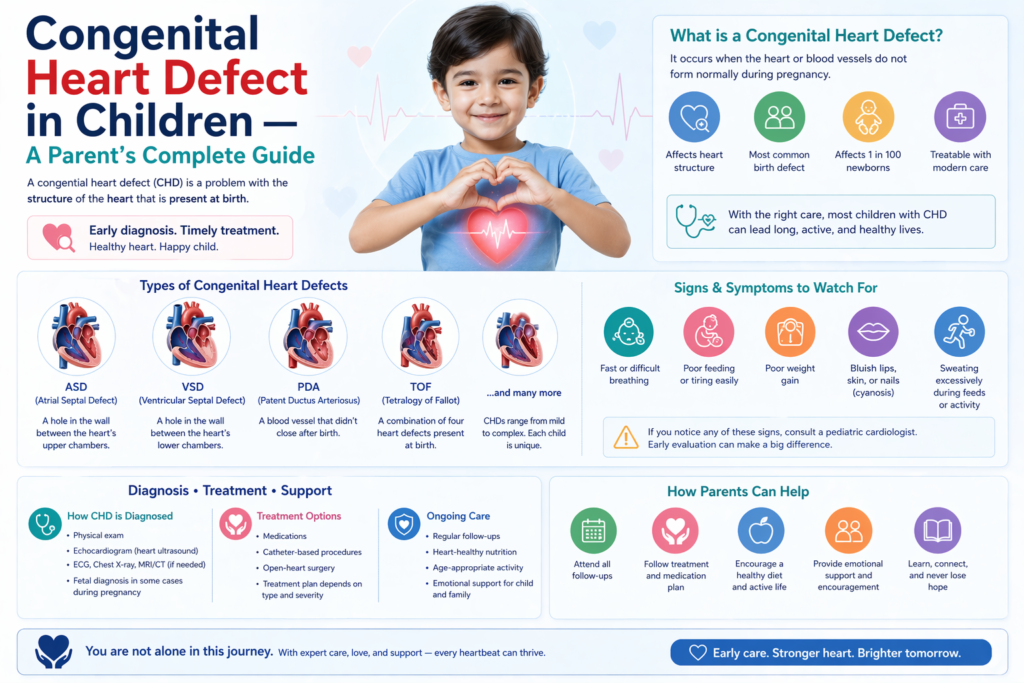
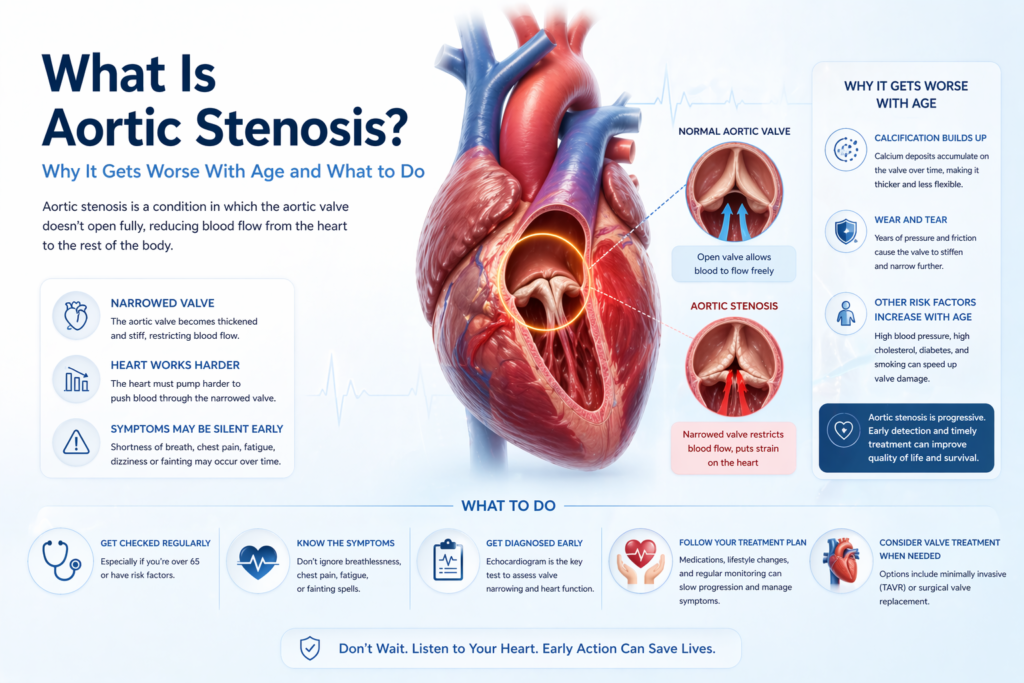
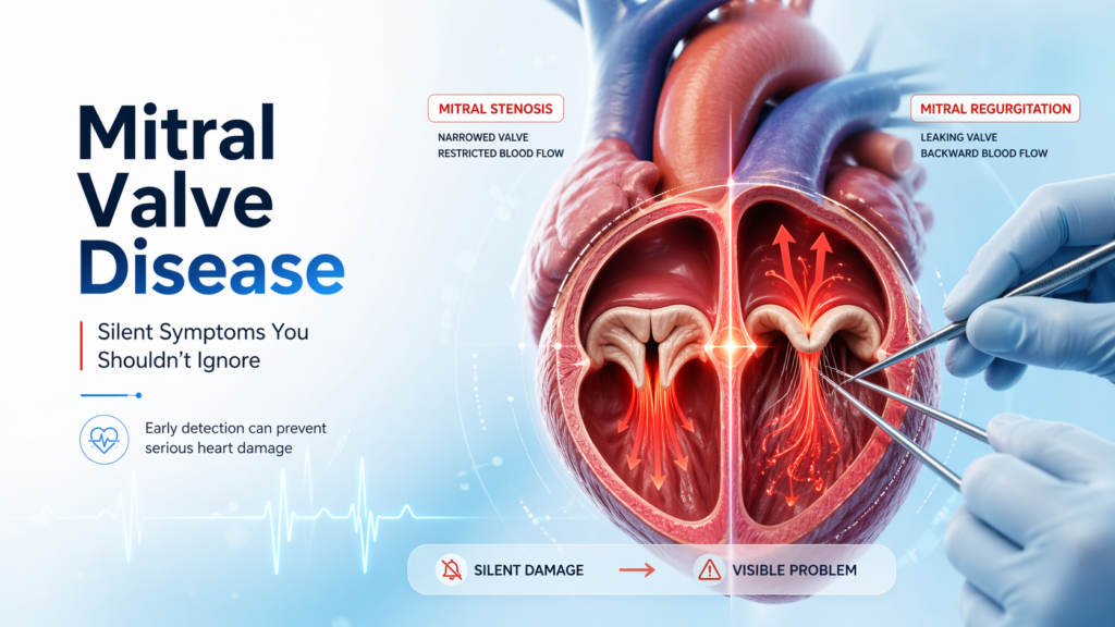
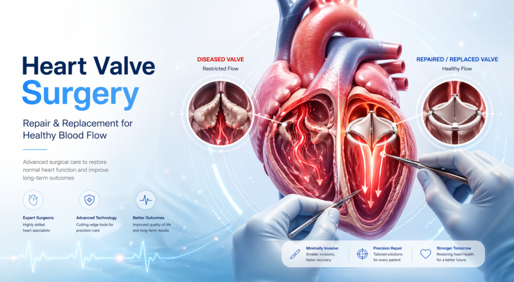
For heart surgery, Dehradun patients have traditionally needed to travel to AIIMS Rishikesh, Delhi, or Chandigarh — journeys of 5–7 hours that are stressful for cardiac patients and costly for families. Dr. Ved Prakash, Director of CTVS at Yatharth Super Speciality Hospitals, Greater Noida — approximately 4 hours from Dehradun via NH-334 and NH-58 — provides full-service cardiac surgery for Dehradun and Uttarakhand patients with the quality of Delhi’s best hospitals and significantly more accessible costs.   Why Dehradun Patients Choose Yatharth Hospital for Heart Surgery Dehradun’s cardiac facilities are limited to cardiology — coronary angiography, angioplasty, and pacemaker implantation are available. But bypass surgery, valve repair and replacement, aortic surgery, and paediatric heart surgery for Dehradun patients require travel. The options are: AIIMS Rishikesh: Excellent institution but long waiting lists for elective CTVS surgery — often 6–12 weeks for non-emergency cases Delhi (AIIMS, Apollo, Max, Fortis): 5–6 hours from Dehradun by road — feasible but exhausting for cardiac patients Yatharth Hospital, Greater Noida: 3.5–4 hours from Dehradun via NH-334/NH-58 — closer than central Delhi, with shorter waiting times and more accessible costs Chandigarh (PGI, Fortis Mohali): 5 hours from Dehradun What Heart Surgery Is Available for Dehradun Patients at Yatharth Hospital? Bypass surgery (CABG): For Dehradun patients referred after angiography — on-pump and off-pump bypass surgery available. No waiting list for referred urgent cases. Mitral and aortic valve surgery: Repair and replacement for rheumatic and degenerative valve disease — significant in the Uttarakhand hill district population TAVI: For elderly Dehradun patients with aortic stenosis too frail for open surgery — available without the journey to AIIMS Delhi Paediatric heart surgery: ASD, VSD, TOF repair for children from Dehradun and Uttarakhand Aortic aneurysm and EVAR/TEVAR Varicose vein laser treatment (EVLT) Getting Started From Dehradun WhatsApp pre-assessment: Send angiogram/echo/CT report to +91-9355255106. Dr. Ved Prakash reviews and advises on urgency, surgical plan, and cost estimate before travel. Online consultation: Available for initial assessment without travel — particularly useful for Dehradun patients to determine urgency before committing to the journey. Insurance: Ayushman Bharat PM-JAY, CGHS, ECHS, and major insurance accepted. Uttarakhand state government health scheme patients should confirm empanelment before visiting. Frequently Asked Questions — Heart Surgery Dehradun How far is Yatharth Hospital from Dehradun? Yatharth Super Speciality Hospitals, Greater Noida is approximately 280–300 km from Dehradun via NH-334 (Haridwar) and NH-58 — a drive of 3.5–4 hours. The route is well maintained and manageable for a family travelling with a cardiac patient. Is bypass surgery available near Dehradun? Full-service CTVS (bypass surgery, valve surgery, aortic surgery) is not available in Dehradun city as of 2026. The nearest cardiac surgery centres are Yatharth Hospital Greater Noida (3.5–4 hours), AIIMS Rishikesh (35 minutes but long waiting lists), and hospitals in Delhi (5–6 hours). Can I get a second opinion for heart surgery from Dehradun without travelling? Yes — Dr. Ved Prakash provides online cardiac second opinion consultations for Dehradun patients. Share your angiogram, echocardiogram, and any clinical reports via WhatsApp at +91-9355255106. A video consultation is arranged and a full clinical opinion is provided before you travel for surgery. Dr. Ved Prakash | Director CTVS — Yatharth Super Speciality Hospitals, Greater Noida 📞 +91-9355255106  | Book Appointment → Tetralogy of Fallot — TOF — is the most common cyanotic congenital heart defect, and tetralogy of Fallot symptoms include the bluish colouration of the skin (cyanosis) that gives this condition its most well-known description: the “blue baby.” With timely surgery, children with tetralogy of Fallot grow up to live full, active lives — but the journey from diagnosis to long-term follow-up requires parents to be well informed. Dr. Ved Prakash, Director of CTVS at Yatharth Super Speciality Hospitals, Greater Noida, explains tetralogy of Fallot symptoms, what surgery involves, and what long-term care is needed.   What Is Tetralogy of Fallot? Tetralogy of Fallot is a combination of four structural heart defects present from birth: A large VSD (Ventricular Septal Defect) — a large hole between the two lower heart chambers Pulmonary stenosis — narrowing of the outflow from the right ventricle to the pulmonary artery (the vessel taking blood to the lungs), sometimes involving the pulmonary valve itself Overriding aorta — the aorta is positioned directly over the VSD, receiving blue (deoxygenated) blood from the right ventricle as well as oxygenated blood from the left Right ventricular hypertrophy — the right ventricle thickens because it is working against the obstructed pulmonary outflow The combination of these four defects means that oxygen-depleted blood bypasses the lungs and is pumped to the body through the aorta — causing the characteristic cyanosis (blue discolouration) that defines tetralogy of Fallot symptoms. Tetralogy of Fallot Symptoms — What Parents Notice Cyanosis (bluish skin): The most characteristic tetralogy of Fallot symptom — blue or purple discolouration of the lips, tongue, fingernails, and toenails. Cyanosis may be present at birth or appear in the first weeks of life as the ductus arteriosus (a fetal blood vessel) closes. “Tet spells” — hypercyanotic episodes: Episodes of sudden, intense cyanosis — the baby turns very blue, becomes irritable or inconsolable, breathes very fast, and may briefly lose consciousness. Tet spells are caused by spasm of the right ventricular outflow tract that suddenly reduces blood flow to the lungs. They are a medical emergency. Squatting in older children: Older children with unrepaired TOF instinctively squat after physical activity — squatting increases systemic vascular resistance and reduces the right-to-left shunting, providing temporary symptom relief Poor growth and feeding difficulties in untreated infants Exercise intolerance in older children with partial defects Clubbing of fingers and toes — in children with long-standing cyanosis How Is Tetralogy of Fallot Diagnosed? Foetal echocardiogram: TOF can often be detected at the 18–20 week anomaly scan — allowing planned delivery at a centre with paediatric cardiac surgery Neonatal echocardiogram: Confirms the diagnosis and anatomical details after birth Pulse oximetry screening: Low oxygen saturation detected at birth routine screening CT cardiac angiography: For detailed assessment of the pulmonary artery anatomy before surgical

Scroll to Top