Uncategorized

varicose vein treatment Ghaziabad
Uncategorized

Varicose Vein Treatment Ghaziabad — EVLT Laser at Yatharth Hospital

  Ghaziabad patients with varicose veins — aching, swollen, or cosmetically troubling — no longer need to travel to central Delhi for laser treatment. EVLT (Endovenous Laser Treatment) is available at Yatharth Super Speciality Hospitals, Greater Noida — 30–40 minutes from Ghaziabad. Walk in. The laser closes the vein. Walk out the same day.   What Is EVLT — In 60 Seconds A fine laser fibre is inserted into the varicose vein through a needle — no incision, no stitches Local anaesthesia (tumescent) numbs the area — no general anaesthesia, no hospital stay The laser seals the vein from inside as the fibre is slowly withdrawn — takes 20–30 minutes Patient walks out. Returns to desk work in 1–2 days. Compression stocking for 2 weeks. Vein closure rate: 90%+ at 5 years For the complete procedure guide, read about how EVLT works. Who Needs Varicose Vein Treatment? Symptom Treatment Urgency Visible bulging veins only — no symptoms Optional (cosmetic) Aching, heaviness, cramping by evening Recommended Leg swelling that does not fully resolve overnight Recommended soon Skin discolouration or hardening around ankle Treat without delay Open wound near ankle that will not heal Urgent — come today Treatment Options at Yatharth Hospital EVLT Laser — first-choice treatment for most varicose veins. Walk-in, walk-out. Radiofrequency Ablation (RFA) — similar to EVLT, slightly different energy type. Same recovery. Sclerotherapy — injection treatment for spider veins and small residual varicosities after EVLT. Surgical stripping — for very large or complex veins not suitable for laser. Under general anaesthesia. Route from Ghaziabad to Yatharth Hospital From Indirapuram / Crossing Republik: 20–25 minutes via NH-58 and Expressway From Ghaziabad city centre: 30–40 minutes via NH-58 From Vaishali / Kaushambi: 25–30 minutes Insurance and Booking Varicose vein treatment is covered by most major health insurance when medically indicated (symptomatic). Cashless facility available. Full varicose vein treatment details on the service page. Call +91-9355255106 to book a vascular surgery OPD appointment A duplex ultrasound scan is done first to map the vein and confirm EVLT suitability EVLT is scheduled — often within days of assessment Frequently Asked Questions — Varicose Vein Treatment Ghaziabad Is varicose vein laser treatment available near Ghaziabad? Yes — EVLT laser treatment at Yatharth Hospital, Greater Noida — 30–40 minutes from Ghaziabad. Walk-in, walk-out, local anaesthesia, 1–2 day return to desk work. What is EVLT and how is it different from surgery? EVLT closes veins from inside via laser through a needle — no incision, no general anaesthesia. Surgical stripping needs incisions and general anaesthesia with 2–3 week recovery. EVLT is now the standard treatment. Does insurance cover varicose vein treatment for Ghaziabad patients? Yes — for symptomatic, medically indicated treatment. Cosmetic-only may not be covered. Confirm with your insurer before booking. Cashless facility available at Yatharth Hospital. Dr. Ved Prakash | Director, CTVS — Yatharth Super Speciality Hospitals, Greater Noida 📞 +91-9355255106  | 📧 drvedprakash@gmail.com  | Book a Consultation →

bypass surgery cost in Delhi NCR
Uncategorized

Bypass Surgery Cost in Delhi NCR 2026 — Complete Patient Guide

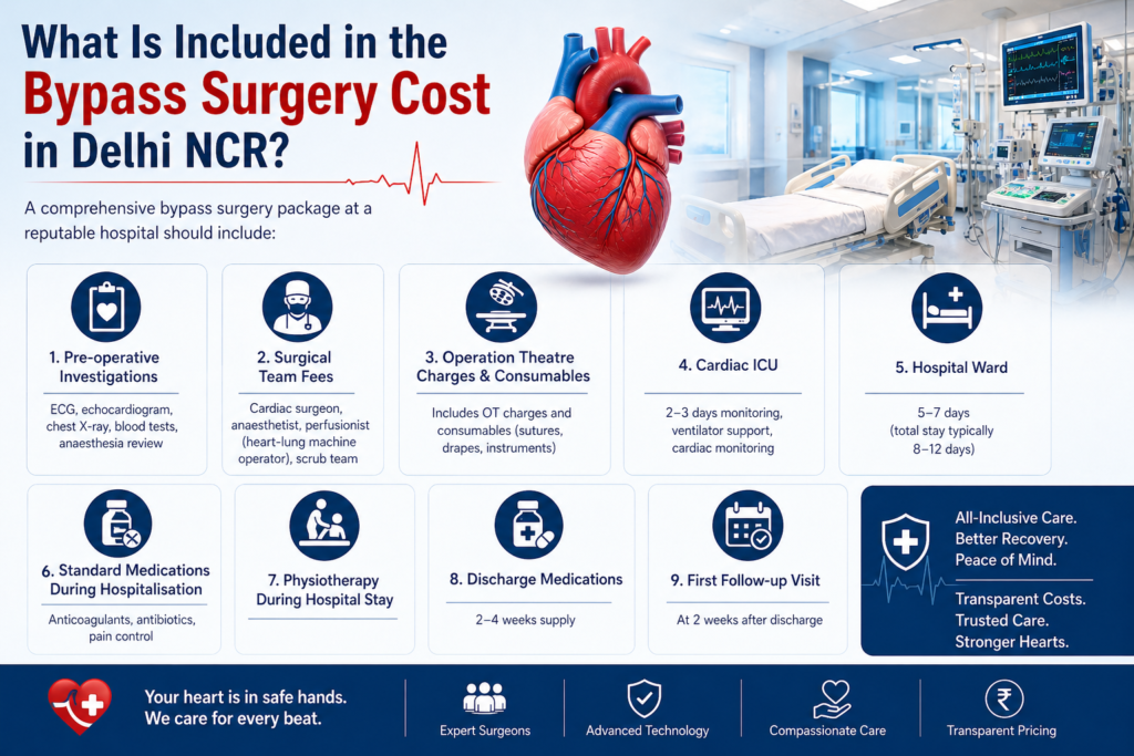
Bypass surgery cost in Delhi NCR is the first question most families ask after an angiogram shows significant blockages — and getting a clear, honest answer is harder than it should be. This guide from Dr. Ved Prakash, Director of CTVS at Yatharth Super Speciality Hospitals, Greater Noida, gives you the complete picture: what bypass surgery cost in Delhi NCR covers, what affects the final bill, how insurance and government schemes work, and what you can do to ensure you receive the best care at the right price.   Bypass Surgery Cost in Delhi NCR — 2026 Price Ranges Bypass surgery cost in Delhi NCR varies based on the hospital category, room type, number of bypass grafts, and patient complexity. As a general guide for 2026: Hospital Category Shared Ward Semi-Private Room Private Room Government hospital (AIIMS, Safdarjung) ₹80,000–1,50,000 ₹1,50,000–2,50,000 ₹2,50,000–3,50,000 Mid-tier private hospital (Yatharth, Sarvodaya, BLK) ₹2,50,000–3,50,000 ₹3,50,000–4,50,000 ₹4,50,000–5,50,000 Premium hospital (Apollo, Max, Fortis Escorts) ₹4,00,000–5,50,000 ₹5,50,000–7,00,000 ₹7,00,000–9,00,000 Note: These are indicative ranges for 2026. Bypass surgery cost in Delhi NCR varies based on individual patient factors — actual quotes should be obtained from the hospital’s billing department after clinical assessment. What Is Included in the Bypass Surgery Cost in Delhi NCR? A comprehensive bypass surgery package at a reputable hospital should include: Pre-operative investigations: ECG, echocardiogram, chest X-ray, blood tests, anaesthesia review Surgical team fees: cardiac surgeon, anaesthetist, perfusionist (heart-lung machine operator), scrub team Operation theatre charges and consumables (sutures, drapes, instruments) Cardiac ICU: 2–3 days monitoring, ventilator support, cardiac monitoring Hospital ward: 5–7 days (total stay typically 8–12 days) Standard medications during hospitalisation: anticoagulants, antibiotics, pain control Physiotherapy during hospital stay Discharge medications: 2–4 weeks supply First follow-up visit at 2 weeks What is often NOT included in the quoted package: coronary angiography before surgery (₹15,000–30,000 separately), CT angiography if needed, blood products if transfusion required, complications or prolonged ICU stay beyond the package duration, and outpatient medications after the first prescription. Factors That Affect Bypass Surgery Cost in Delhi NCR Number of bypass grafts: Single bypass costs less than triple bypass — more grafts mean longer operative time and more graft material On-pump vs off-pump: Off-pump (OPCAB) surgery typically adds 10–15% to the cost due to the specialised stabiliser equipment Arterial grafts used: Total arterial revascularisation (LIMA + radial artery) may add to cost but improves long-term durability Patient complexity: Emergency surgery, redo bypass (after previous surgery), low ejection fraction, or concurrent valve surgery — all increase cost ICU duration: Patients with complications who stay in ICU longer than the package duration incur additional daily ICU charges (₹8,000–20,000/day depending on hospital) Room category: The single largest variable in bypass surgery cost in Delhi NCR — private room vs shared ward can differ by ₹50,000–1,00,000 for the same surgery Insurance Coverage for Bypass Surgery Cost in Delhi NCR Most comprehensive health insurance policies cover bypass surgery cost in Delhi NCR as an inpatient procedure. Key points: Cashless treatment: Available at empanelled hospitals. Submit pre-authorisation to your TPA (Third Party Administrator) 2–3 days before elective surgery with the angiogram report and surgeon’s recommendation letter. Most private hospitals including Yatharth Hospital have dedicated insurance facilitation teams. Waiting period: Most policies have a 1–4 year waiting period for pre-existing conditions. If bypass surgery is needed urgently (within the waiting period), claim reimbursement after hospital discharge — insurers cannot deny medically necessary emergency surgery. Sub-limits: Some older policies have sub-limits on ICU charges or surgeon fees — check your policy schedule before finalising the hospital. Top-up policies: If your current sum insured is below ₹5 lakhs and bypass surgery cost in Delhi NCR exceeds this, a top-up policy bridges the gap — worth considering for every cardiac patient. Government Schemes Covering Bypass Surgery Cost in Delhi NCR Ayushman Bharat PM-JAY: Covers bypass surgery cost in Delhi NCR up to ₹5 lakhs per family per year for eligible beneficiaries (below poverty line / specific government categories). Yatharth Hospital is empanelled. Check eligibility at pmjay.gov.in. CGHS (Central Government Health Scheme): Covers central government employees and pensioners. Bypass surgery cost in Delhi NCR is reimbursed at CGHS package rates — which may be lower than private rates. Empanelment status of hospital matters. ECHS (Ex-Servicemen Contributory Health Scheme): Similar to CGHS for defence personnel. Yatharth Hospital Greater Noida accepts ECHS cards. State government schemes: UP government’s Mukhyamantri Jan Arogya Yojana covers eligible UP residents for bypass surgery cost at empanelled hospitals in Greater Noida. How to Reduce Bypass Surgery Cost in Delhi NCR Legitimately Choose a shared ward or semi-private room — the surgery is identical, only the room differs Ask for a detailed package quote in writing before admission — including what extras will be charged Ensure pre-authorisation is obtained from your insurer before the surgery date Do not delay surgery while financial arrangements are made — deterioration may convert an elective surgery to an emergency, significantly increasing cost and risk Ask specifically whether the surgeon who quoted your case will personally perform your surgery Bypass Surgery Cost at Yatharth Super Speciality Hospitals, Greater Noida Bypass surgery cost in Delhi NCR at Yatharth Super Speciality Hospitals, Greater Noida is in the mid-tier range — offering the surgical expertise of Dr. Ved Prakash (8+ years experience including Medanta, Narayana, and Sarvodaya) in a NABH-accredited hospital at a price point accessible to UP and Delhi NCR patients without requiring travel to central Delhi. Cashless insurance, Ayushman Bharat, CGHS, ECHS, and corporate tie-ups are available. For a specific quote for your case, share your angiogram report via WhatsApp or book a bypass surgery consultation in Delhi NCR. Frequently Asked Questions — Bypass Surgery Cost in Delhi NCR What is the average bypass surgery cost in Delhi NCR in 2026? The average bypass surgery cost in Delhi NCR in 2026 is ₹3,50,000–4,50,000 in a semi-private room at a reputable mid-tier private hospital. At premium hospitals (Apollo, Max, Fortis), bypass surgery cost in Delhi NCR ranges ₹5,50,000–8,00,000 for comparable surgery in a private room. Is bypass surgery cost

diet after heart surgery India
Uncategorized

diet after heart surgery India— What to Eat and What to Avoid

Diet after heart surgery in India is one of the most common concerns patients ask about after bypass or valve surgery — and unfortunately, the advice many patients receive is so vague (“eat healthy”) that it is practically useless. Dr. Ved Prakash, Director of CTVS at Yatharth Super Speciality Hospitals, Greater Noida, provides a complete, India-specific diet guide after heart surgery — covering the foods to eat, the foods to avoid, and honest guidance on how to navigate desi cooking in the recovery period.   Why Diet After Heart Surgery in India Matters More Than Most Patients Realise Diet after heart surgery in India directly affects: The lifespan of bypass grafts and stents — a high-fat diet accelerates new plaque formation in the grafted vessels Blood cholesterol and triglyceride levels — which must be controlled with both medication AND diet Blood pressure control — excessive sodium intake raises blood pressure and stresses the repaired heart Blood sugar management — particularly important since many bypass surgery patients have diabetes Wound healing and recovery — adequate protein is essential in the 6–8 weeks after surgery The diet after heart surgery in India is not about punishment or deprivation — it is about making smart, sustainable choices that protect your surgical investment for the next 15–20 years. What to Eat After Heart Surgery in India Grains and Carbohydrates Best choices: Whole wheat roti (2-3 per meal), brown rice in moderate portions, jowar roti, bajra roti, oats, daliya (broken wheat porridge) Avoid: White rice in large quantities (high glycaemic index — particularly problematic in diabetic patients), maida (refined flour) products — puri, paratha, naan, bread Portion: 2–3 rotis per meal or half a katori of cooked brown rice. Not an unlimited staple. Protein — Critical for Recovery Best vegetarian choices: Dal (moong, masoor, chana — all excellent) 1–2 katori per day; low-fat paneer in moderate amounts; curd (dahi — plain, not sweetened); sprouts; soya chunks; egg whites Non-vegetarian: Grilled or boiled chicken breast (without skin); fish — especially rohu, katla, sardine (omega-3 rich); egg whites Avoid: Red meat (mutton, beef), full-fat paneer in large quantities, fried proteins Fruits and Vegetables No meaningful restriction on vegetables — include 3–4 varieties daily, especially green leafy vegetables (spinach, methi, palak), tomatoes, onions, garlic, and cruciferous vegetables Fruits: 2 portions daily — guava, apple, papaya, berries, orange, amla (excellent vitamin C for wound healing). Avoid: mango, banana, chikoo in large quantities if diabetic — high sugar content. Important: If taking warfarin (blood thinner after mechanical valve surgery), do not suddenly increase or decrease your intake of green leafy vegetables — maintain a consistent, moderate amount and inform your doctor Fats and Oils in Indian Cooking This is the area where patients make the most mistakes. The diet after heart surgery in India does not mean zero oil — it means choosing the right oils and using them in appropriate quantities. Best oils: Mustard oil, olive oil, groundnut oil, rice bran oil — used in limited quantity (2–3 teaspoons per day total) Avoid: Vanaspati (dalda), coconut oil (in large amounts), palm oil — high in saturated fat Ghee: Small amounts of homemade ghee (half a teaspoon 3–4 times per week) are acceptable for most patients — the data on pure ghee in moderation in Indians is less alarming than its reputation suggests. Commercially produced ghee or large amounts of ghee daily should be avoided. Going completely oil-free is wrong — fats are necessary for fat-soluble vitamin absorption (A, D, E, K), brain function, and satiety. Eliminating fat entirely often leads to compensating with more refined carbohydrates — which is worse for the heart. Heart-Protective Foods to Add Daily Garlic: 2–3 raw or cooked cloves daily — modest LDL-lowering and anti-platelet effect Flaxseeds (alsi): 1 tablespoon ground daily — excellent plant omega-3 source Walnuts: 4–5 walnuts daily — omega-3, vitamin E, and anti-inflammatory Amla (Indian gooseberry): 1 fresh amla or 1 teaspoon amla powder daily — exceptionally high vitamin C Methi (fenugreek seeds): Soaked overnight, consumed in the morning — helps blood sugar and cholesterol control What to Avoid After Heart Surgery in India Namkeen, papad, pickle, processed snacks: Extremely high in sodium — significantly raises blood pressure Restaurant and dhaba food: Cooked in large quantities of oil, high sodium, unknown quality fats — avoid for the first 6 months at minimum Full cream milk and curd: Switch to low-fat (toned) milk and curd Fried foods of any kind: Puri, bhatura, samosa, pakora, chips — avoid entirely for 12 months after surgery Mithai and sweets: High in sugar and saturated fat — particularly dangerous for diabetic bypass patients. Festivals are not an excuse to make exceptions. Alcohol: Avoid completely for at least 3 months after surgery. After that, if the cardiologist permits — maximum 1 small drink per day for men, occasionally. Avoid entirely if on warfarin. Diet After Heart Surgery India — The Practical Weekly Framework Meal Good Choices Early morning Soaked methi seeds + 4 walnuts + amla Breakfast Oats with low-fat milk, or daliya, or 2 egg whites with whole wheat toast Lunch 2 whole wheat rotis + dal (1 katori) + sabzi (cooked in minimal oil) + salad Afternoon Fruit (guava/apple) + plain low-fat dahi Dinner 2 rotis + light sabzi or grilled fish/chicken + dal or sprouts Bedtime Half katori low-fat milk (plain, no sugar) Frequently Asked Questions — Diet After Heart Surgery India Can I eat dal roti after bypass surgery? Yes — whole wheat roti and dal are excellent choices for the diet after heart surgery in India. Dal provides plant protein, fibre, and complex carbohydrates with a low glycaemic index. Use minimal oil in the tadka. Avoid high-sodium spice mixes and pickle on the side. Can I eat ghee after bypass surgery? Small amounts of homemade ghee — half a teaspoon occasionally — are acceptable for most patients as part of a balanced diet after heart surgery in India. Daily or large-quantity ghee should be avoided. Commercially produced ghee contains more

what is aortic aneurysm
Uncategorized

what is aortic aneurysm? Silent but Dangerous — What You Need to Know

An aortic aneurysm is a bulge or ballooning in the wall of the aorta — the body’s main artery — and it is one of the most dangerous conditions in medicine because an aortic aneurysm typically causes no symptoms until it is on the verge of rupturing. Dr. Ved Prakash, Director of CTVS at Yatharth Super Speciality Hospitals, Greater Noida, explains what is an aortic aneurysm, when it becomes dangerous, how it is diagnosed, and what surgery involves.   What Is an Aortic Aneurysm? The aorta is the largest artery in the body — rising from the heart, arching through the chest, and running down through the abdomen to supply the entire body. The normal aortic diameter is approximately 2.5–3.0 cm. An aortic aneurysm is an abnormal widening of the aorta to more than 1.5 times its normal diameter — typically defined as greater than 3.0 cm in the abdominal aorta or greater than 4.5 cm in the thoracic (chest) aorta. As an aortic aneurysm enlarges, the wall of the aorta becomes progressively thinner and weaker. At a critical size, the wall can rupture — releasing enormous volumes of blood into the chest or abdomen. Rupture of an aortic aneurysm carries a mortality rate exceeding 80% even with emergency surgery. This is why detecting and treating an aortic aneurysm before rupture is so critical. Types of Aortic Aneurysm Abdominal Aortic Aneurysm (AAA) An aortic aneurysm in the portion of the aorta that runs through the abdomen — the most common location. AAAs occur primarily in men over 65 with a history of smoking, hypertension, or a family history of aortic aneurysm. They are frequently detected incidentally on ultrasound or CT scan performed for another reason — because they rarely cause symptoms until they are very large or rupturing. Thoracic Aortic Aneurysm (TAA) An aortic aneurysm in the aorta within the chest — either the ascending aorta (rising from the heart), the arch (the curved section at the top), or the descending aorta (running down behind the heart). TAAs are more often associated with genetic conditions (Marfan syndrome, bicuspid aortic valve) and hypertension, and can occur at younger ages than AAAs. What Causes an Aortic Aneurysm? Atherosclerosis: The primary cause of AAA — decades of high blood pressure, high cholesterol, and smoking cause the aortic wall to degenerate Hypertension: The most important modifiable risk factor — controlling blood pressure slows aortic aneurysm growth Smoking: Doubles aortic aneurysm risk and accelerates growth rate significantly Genetic connective tissue disorders: Marfan syndrome, Loeys-Dietz syndrome, and Ehlers-Danlos syndrome predispose to thoracic aortic aneurysms — often at young ages Bicuspid aortic valve: Associated with progressive thoracic aortic aneurysm — regular aortic surveillance with echocardiogram or CT is essential Family history: First-degree relatives of AAA patients have a 10–15% lifetime risk — screening ultrasound is recommended Aortic Aneurysm Symptoms — Why It Is Called the Silent Killer The vast majority of aortic aneurysms are completely asymptomatic until rupture or acute expansion. When symptoms do occur, they are usually: Deep, constant back or abdominal pain — particularly in the lower back, which may be confused with musculoskeletal pain. This is often a sign of a rapidly expanding or leaking AAA — a surgical emergency. Pulsating sensation in the abdomen — a pulsatile mass felt in the centre of the abdomen, like a second heartbeat. Most often felt by the patient themselves or detected on examination. Chest or back pain in thoracic aneurysm — from pressure on surrounding structures. A descending thoracic aneurysm may cause hoarseness (from recurrent laryngeal nerve compression) or difficulty swallowing. Rupture: Sudden, severe abdominal or back pain, collapse, and circulatory shock. A ruptured aortic aneurysm is the surgical emergency that every vascular surgeon dreads — and that mortality figures make sobering reading. When Does an Aortic Aneurysm Need Surgery? The treatment decision for an aortic aneurysm is based primarily on size — because larger aneurysms carry a significantly higher annual rupture risk: AAA Diameter Annual Rupture Risk Recommendation 3.0–4.4 cm <0.5% Surveillance ultrasound every 12 months 4.5–5.0 cm 1–3% Surveillance every 6 months + surgical review 5.0–5.5 cm 5–10% Surgery recommended for most patients Over 5.5 cm 10–25% Surgery urgently recommended Any size — rapidly expanding (>1cm/year) High Surgery recommended regardless of size Aortic Aneurysm Treatment — EVAR vs Open Surgery EVAR (Endovascular Aneurysm Repair): A stent-graft is delivered through the femoral arteries in the groin and positioned inside the aneurysm — excluding it from the circulation without opening the abdomen. Recovery is significantly faster (2–3 days hospital stay) and surgical risk is lower. Suitable for most infrarenal AAAs with suitable anatomy. Open surgical repair: The aneurysm is exposed through an abdominal incision, clamped, and replaced with a synthetic (Dacron) graft. More invasive — 7–10 day hospital stay — but the definitive lifelong repair. Required when the anatomy is not suitable for EVAR, or for juxtarenal/suprarenal aneurysms. TEVAR (Thoracic Endovascular Aortic Repair): The endovascular equivalent of EVAR for descending thoracic aortic aneurysms — a stent-graft deployed through the femoral artery to reline the thoracic aorta. Frequently Asked Questions — What Is Aortic Aneurysm What is an aortic aneurysm and is it always fatal? An aortic aneurysm is an abnormal bulging of the aorta. It is not immediately fatal — most detected aneurysms are small, grow slowly, and are treated safely with elective surgery before they rupture. A ruptured aortic aneurysm, however, carries a mortality rate exceeding 80%. Early detection and timely elective repair are what save lives. How is an aortic aneurysm found if there are no symptoms? Most aortic aneurysms are discovered incidentally — on an ultrasound for kidney or liver problems, or on a CT scan. Screening ultrasound is recommended for all men over 65 with a history of smoking, and for first-degree relatives of AAA patients. Can an aortic aneurysm be treated without surgery? Small aneurysms under 5 cm are managed with surveillance (regular size monitoring), blood pressure control, statin therapy, and smoking cessation — which slow growth. No medication reduces

Coronary Artery Disease
Uncategorized

Coronary Artery Disease — Stages, Symptoms and When Surgery Is Needed

Coronary artery disease is not a single event — it is a decades-long process that progresses through identifiable stages, with each stage carrying different symptoms, different risks, and different treatment implications. Understanding where you or a family member sits in this progression is the most important step toward making the right treatment decision. Dr. Ved Prakash, Director of CTVS at Yatharth Super Speciality Hospitals, Greater Noida, explains coronary artery disease stages and symptoms, and when each stage crosses the threshold for surgical intervention.   What Is Coronary Artery Disease? Coronary artery disease (CAD) is the buildup of atherosclerotic plaque — a mixture of cholesterol, inflammatory cells, calcium, and fibrous tissue — inside the walls of the coronary arteries that supply the heart muscle with blood. As this plaque accumulates over years, it progressively narrows the artery lumen. When narrowing reaches approximately 70%, blood flow during exertion is insufficient and symptoms begin. When a plaque ruptures and a clot forms suddenly, the result is a heart attack. India has one of the highest burdens of coronary artery disease globally — affecting patients 10–15 years younger on average than in Western countries, and with a disproportionately high rate of multi-vessel disease at first presentation. Coronary Artery Disease Stages — The Full Progression Stage 1 — Subclinical Atherosclerosis (No Symptoms) Plaque begins accumulating in the coronary artery walls from as early as the 20s and 30s in high-risk individuals — those with diabetes, hypertension, high cholesterol, a strong family history, or heavy smoking history. At this stage, the narrowing is less than 50% and blood flow is not restricted. There are no symptoms whatsoever. This stage is detectable with CT coronary calcium scoring and CT coronary angiography — tests that show plaque burden before any symptoms develop. This is the stage where lifestyle modification and statin therapy are most effective at slowing or halting progression. Stage 2 — Stable Angina (Predictable Chest Pain on Exertion) When the coronary artery narrows beyond 70%, blood flow during physical exertion becomes insufficient for the heart muscle’s increased demand. The result is stable angina — chest pain or tightness that comes on predictably after a certain amount of exertion and resolves completely within 5–10 minutes of rest. Patients describe stable angina as: central chest tightness, pressure, heaviness, or aching — sometimes radiating to the left arm, jaw, or between the shoulder blades. The pain is consistent — the same exertion produces the same symptoms. It does not occur at rest. If you develop this symptom pattern, see a cardiologist immediately for an ECG, stress test, and likely coronary angiography. Diabetic patients and women frequently do not experience typical chest pain — they present with breathlessness, jaw pain, left arm pain, or unexplained fatigue as their angina equivalent. These atypical presentations lead to delayed diagnosis. Stage 3 — Unstable Angina / NSTEMI (Plaque Rupture) When atherosclerotic plaque becomes unstable — thin-capped, inflamed, and vulnerable — it can rupture. A ruptured plaque triggers immediate clot formation (thrombus) at the rupture site. If the clot partially blocks the artery, blood flow at rest is compromised. The result is: Unstable angina: Chest pain at rest, or with minimal exertion, or that is getting progressively more frequent and severe. A dangerous change from a previously stable pattern. NSTEMI (Non-ST Elevation Myocardial Infarction): Partial blockage with some heart muscle damage — troponin rises on blood tests but the ECG does not show the classic ST elevation of a full heart attack. Both unstable angina and NSTEMI require hospital admission and urgent coronary angiography — typically within 24–48 hours. This is not a situation where a patient should wait for a scheduled appointment. Stage 4 — STEMI (Complete Heart Attack) When the clot at the rupture site completely occludes the artery, blood supply to the downstream heart muscle stops entirely. This is a STEMI — ST-Elevation Myocardial Infarction — a full heart attack. Every minute of complete occlusion destroys heart muscle that will never recover. Time from symptom onset to opening the blocked artery is the single most important determinant of how much heart muscle is saved. STEMI symptoms: Sudden severe chest pain at rest — crushing, squeezing, or pressure sensation. May radiate to the left arm, jaw, or back. Often accompanied by sweating, nausea, breathlessness, and a feeling of impending doom. Can also present without chest pain in diabetics — only breathlessness and sweating. STEMI is a medical emergency. Call emergency services immediately. Do not drive yourself to hospital. How Many Arteries Are Blocked — And Why It Matters Coronary artery disease is classified by how many vessels are affected: Vessel Count Definition Typical Treatment Single vessel One artery with significant blockage Angioplasty usually appropriate Two vessel (double vessel) Two arteries significantly blocked Heart Team decision — depends on location and patient profile Three vessel (triple vessel) All three main arteries blocked Bypass surgery strongly preferred Left main disease Trunk artery before LAD and LCx Bypass surgery in most cases Risk Factors That Accelerate Coronary Artery Disease Diabetes — the single most aggressive accelerator of CAD in Indian patients. Doubles the risk and significantly worsens outcomes after heart attack. Hypertension — damages the arterial endothelium, accelerating plaque deposition Smoking — directly toxic to coronary endothelium; smokers develop CAD 10 years earlier than non-smokers High LDL cholesterol — primary driver of plaque accumulation; statin therapy is the cornerstone of prevention Family history — first-degree relative with heart disease before 55 (men) or 65 (women) doubles your risk Sedentary lifestyle and central obesity — particularly the android fat distribution pattern common in South Asians When Coronary Artery Disease Requires Surgery Not all coronary artery disease requires surgery. The threshold is determined by: Symptoms that are not adequately controlled with medication Anatomical findings on angiography that predict a survival benefit from surgery — triple vessel disease, left main disease, or high SYNTAX score Ejection fraction below 35% with multi-vessel disease — where complete revascularisation by surgery improves both symptoms and survival The decision between bypass

heart valve surgery cost in Delhi
Uncategorized

What Is TAVI? Heart Valve Replacement Without Open-Chest Surgery

TAVI — Transcatheter Aortic Valve Implantation — is a procedure that replaces the aortic heart valve through a catheter inserted through the leg artery, without opening the chest at all. It has transformed cardiac care for thousands of patients who were previously told they were too old or too unwell for heart surgery. Dr. Ved Prakash, Director of CTVS at Yatharth Super Speciality Hospitals, Greater Noida, explains the TAVI procedure, who qualifies, and how the decision between TAVI and open surgery is made.   What Is TAVI — The Basics TAVI (also called TAVR — Transcatheter Aortic Valve Replacement) is a catheter-based procedure that delivers a replacement aortic valve to the heart through an artery in the groin (femoral artery) — or sometimes through the chest wall (transapical) — without making a large chest incision or stopping the heart. The new biological tissue valve is compressed onto a small expandable frame and mounted on a catheter. Once positioned precisely inside the diseased native aortic valve using X-ray and echocardiographic guidance, the new valve is deployed — opening like a flower and immediately taking over the function of the diseased valve. The old, calcified valve leaflets are pushed aside. The procedure takes 1–2 hours. Most patients walk the next day and go home within 3–5 days. Why Was the TAVI Procedure Developed? Aortic stenosis — the narrowing of the aortic valve — is the most common serious valve disease in patients over 65. Untreated severe aortic stenosis carries an average survival of 1–3 years once symptoms appear. For decades, the only treatment was open-heart surgical valve replacement (SAVR) — which in elderly or medically frail patients carries significant risk. The TAVI procedure was developed specifically to offer these high-risk patients a life-saving valve replacement without the trauma of open surgery. First performed in 2002, TAVI has now been performed on over 1 million patients worldwide — with outcomes equivalent to surgery in high and intermediate-risk patients, and now expanding to low-risk patients as well. Who Is a Candidate for the TAVI Procedure? The TAVI procedure is recommended for patients with: Severe symptomatic aortic stenosis — confirmed on echocardiogram (valve area <1.0 cm², mean gradient >40 mmHg) AND symptoms of breathlessness, chest pain, or syncope High or intermediate surgical risk — assessed using the STS Score or EuroSCORE II (heart surgery risk calculators) Suitable aortic valve anatomy and femoral artery access — confirmed by CT angiography before the TAVI procedure The TAVI procedure is also being used increasingly in lower-risk patients — and as of the most recent ESC/ACC guidelines (2024), TAVI is a Class I recommendation across all risk groups for severe symptomatic aortic stenosis in patients over 75. TAVI vs Open Heart Surgery (SAVR) — How Is the Decision Made? This is where the Heart Team concept is essential — and where most TAVI articles fail patients by not explaining it. Every patient considered for aortic valve replacement at Yatharth Hospital is evaluated by a Heart Team — consisting of Dr. Ved Prakash (cardiac surgeon) + an interventional cardiologist + an anaesthetist — who review the echocardiogram, CT angiography, and clinical status together before recommending TAVI or surgery. Factor Favour TAVI Favour Open Surgery (SAVR) Age Over 75 years Under 65–70 years Surgical risk High or intermediate Low (young, fit patient) Lung function Poor (COPD, pulmonary fibrosis) Normal Aortic calcification Severe (avoids aortic clamping) Minimal Need for simultaneous procedures No — valve only Yes — coronary bypass + valve Valve durability need Less critical (older patient) Long term (younger patient) What Happens During the TAVI Procedure — Step by Step Anaesthesia: General anaesthesia or conscious sedation (local anaesthesia + sedation) — depending on the patient and the centre’s protocol Access: A small puncture in the femoral artery in the groin. No chest incision in transfemoral TAVI. Crossing the valve: A guidewire is advanced through the femoral artery, up the aorta, and across the diseased aortic valve — guided by X-ray Balloon valvuloplasty: A small balloon briefly opens the calcified valve to create space for the new valve Valve deployment: The replacement valve on its delivery system is positioned precisely at the level of the native aortic valve — then deployed. The valve immediately starts functioning. Confirmation: Echocardiography and angiography confirm the valve is working correctly, there is no significant leakage, and the coronary arteries remain open Access closure: The femoral artery puncture is closed with a vascular closure device — no surgical cut-down needed in most cases Life After the TAVI Procedure — What to Expect Hospital stay: 3–5 days in most patients — significantly shorter than the 7–10 days after open surgery Walking: The next day after the TAVI procedure in most cases Medications after TAVI: Aspirin lifelong (to prevent clotting on the new valve) + clopidogrel for 3–6 months. Warfarin is generally NOT required after TAVI unless you also have atrial fibrillation. Pacemaker: Approximately 10–15% of TAVI patients require a permanent pacemaker — the new valve can sometimes interfere with the heart’s electrical conduction system. This is discussed with every patient before the TAVI procedure. Echocardiogram follow-up: At 1 month, 1 year, and annually thereafter — to check valve function and detect any late complications TAVI valve lifespan: 10–15 years in most patients. If a TAVI valve wears out, a second TAVI (valve-in-valve) can often be performed. Frequently Asked Questions — What Is TAVI Procedure What is the TAVI procedure and is it major surgery? The TAVI procedure is a minimally invasive catheter-based aortic valve replacement — not open-heart surgery. No chest incision is made, the heart is not stopped, and no heart-lung machine is used. Most patients are discharged in 3–5 days and return to full activity within 2–4 weeks. Is TAVI safe for elderly patients? Yes — TAVI was specifically developed for high-risk and elderly patients who cannot safely undergo open surgery. In patients over 80 with severe aortic stenosis and multiple comorbidities, the TAVI procedure offers a survival benefit and quality-of-life improvement that would

peripheral artery disease symptoms
Uncategorized

Peripheral Artery Disease Symptoms — Why Leg Pain While Walking Is a Warning Sign

Peripheral artery disease symptoms are one of the most under-recognised warning signs in Indian medicine — and in diabetic patients, peripheral artery disease symptoms are particularly dangerous because they are often absent entirely until the disease is advanced. Dr. Ved Prakash, Director of CTVS and Vascular Surgery at Yatharth Super Speciality Hospitals, Greater Noida, explains peripheral artery disease symptoms, causes, risk groups, and when surgery or endovascular treatment is needed in Delhi NCR.   What Is Peripheral Artery Disease? Peripheral artery disease (PAD) is atherosclerosis of the arteries supplying the legs — the same process of plaque buildup that causes coronary artery disease in the heart, but affecting the vessels below the waist. As plaque builds up, the leg arteries narrow — reducing blood supply to the muscles and tissues of the leg. The result is the characteristic peripheral artery disease symptom of claudication: leg pain or cramping that comes on with walking and is relieved within 5 minutes of rest. Peripheral artery disease affects approximately 200 million people worldwide and is significantly underdiagnosed in India — particularly in patients with diabetes, where the classic peripheral artery disease symptoms may be absent due to accompanying nerve damage (neuropathy). Peripheral Artery Disease Symptoms — The Spectrum from Mild to Critical Stage 1 — Claudication (Intermittent Leg Pain on Walking) The hallmark peripheral artery disease symptom. Cramping, tightness, or aching pain in the calf, thigh, or buttock that begins predictably after a certain distance of walking — the claudication distance. The pain forces the patient to stop. After 5–10 minutes of rest, it completely resolves — and walking resumes. If you can walk 500 metres before the pain stops you, claudication is mild. If 100 metres stops you, claudication is severe. The location of the pain indicates the level of arterial disease: calf claudication means disease in the superficial femoral artery (mid-thigh level); thigh and buttock claudication indicates disease higher up, in the iliac arteries of the pelvis. Stage 2 — Reduced Claudication Distance (Disease Progression) As peripheral artery disease progresses, the claudication distance shortens. A patient who could walk 300 metres 2 years ago can now manage only 100 metres. This reflects worsening arterial narrowing — and is a signal that intervention should be considered before the next stage develops. Stage 3 — Rest Pain (Critical Ischaemia) When peripheral artery disease becomes severe enough, the foot no longer receives adequate blood even at rest. The patient experiences burning pain in the foot — classically worse at night, often requiring the foot to be hung over the edge of the bed (which uses gravity to increase blood flow). Rest pain is a medical emergency — limb loss occurs within weeks if not treated. Stage 4 — Tissue Loss (Gangrene or Ulceration) The most advanced peripheral artery disease symptom — wounds that do not heal, or areas of blackening (gangrene) on the toes or foot. In diabetic patients, a small foot wound that does not heal despite weeks of dressing is often the first presentation of severe underlying PAD. This stage requires urgent vascular surgery assessment — delay means amputation. Peripheral Artery Disease Symptoms in Diabetic Patients — The Silent Crisis Diabetic neuropathy damages the peripheral nerves — meaning diabetic patients with severe PAD may have no pain at all. They do not experience claudication, no rest pain, and they may not feel a wound developing on their foot. The first sign of advanced peripheral artery disease in a diabetic patient is often a non-healing ulcer or blackened toe — by which time the disease is already critical. All diabetic patients over 50 should have an annual ABI (Ankle-Brachial Index) measurement — a simple, painless test that compares blood pressure in the ankle to the arm to screen for peripheral artery disease silently. What Causes Peripheral Artery Disease? Peripheral artery disease is caused by atherosclerosis — the same risk factors as heart disease: Smoking: The single strongest modifiable risk factor for PAD — smokers have 3–5 times the peripheral artery disease risk of non-smokers. Stopping smoking slows PAD progression significantly. Diabetes: Doubles PAD risk and dramatically worsens outcomes — particularly for below-knee arterial disease Hypertension (high blood pressure) High cholesterol (hyperlipidaemia) Family history of vascular disease Obesity and sedentary lifestyle How Is Peripheral Artery Disease Diagnosed? ABI (Ankle-Brachial Index): Non-invasive, painless. A ratio below 0.9 confirms significant PAD. Below 0.5 indicates critical ischaemia. Duplex Doppler ultrasound: Maps the arterial anatomy and identifies the location and severity of narrowings CT angiography: Detailed 3D imaging of the arteries from abdomen to foot — essential for planning endovascular or surgical intervention Conventional angiography: Catheter-based — performed in the interventional suite when treatment (angioplasty or bypass) is planned immediately Peripheral Artery Disease Treatment in Delhi NCR Medical management: Antiplatelet drugs (aspirin or clopidogrel), statin therapy, blood pressure control, diabetes management, supervised exercise program (increases claudication distance by 50–100%), and strict smoking cessation Endovascular treatment (angioplasty/stenting): For suitable blockages — a balloon catheter opens the narrowed artery and a stent is placed to keep it open. Minimally invasive, no incision, 1–2 day hospital stay. Peripheral bypass surgery: For long or multiple blockages not suitable for endovascular treatment — a graft (either synthetic or vein graft from the leg) is used to bypass the blocked section. Effective and durable, with recovery of 4–6 weeks. For patients with critical ischaemia, urgent peripheral vascular disease treatment in Delhi NCR is available at Yatharth Super Speciality Hospitals, Greater Noida.

Uncategorized

What Is TAVI? Aortic Valve Replacement Without Opening the Chest

Aortic Valve Replacement Without Opening the Chest: TAVI — Transcatheter Aortic Valve Implantation — is a procedure that replaces a diseased aortic heart valve through a catheter passed through the leg artery, without any chest incision and without stopping the heart. For the millions of elderly patients in India who have been told their aortic valve is severely narrowed but that open surgery is too risky at their age, TAVI is often the answer. Dr. Ved Prakash, Director of CTVS at Yatharth Super Speciality Hospitals, Greater Noida, explains what TAVI is, how the procedure works, and crucially, how the decision between TAVI and open surgery is made. Why Was TAVI Developed? Aortic stenosis — narrowing of the aortic valve — is the most common serious valve disease in patients over 65. Once symptoms appear, the average untreated survival is 1–3 years. For decades, the only treatment was open surgical valve replacement, which carries significant risk in frail, elderly, or medically complex patients. The TAVI procedure was developed specifically to treat this group — offering a life-saving valve replacement without the trauma of open surgery. First performed in humans in 2002, TAVI has now been performed on over a million patients worldwide with results that equal or exceed open surgery in high-risk groups. What Is Aortic Stenosis — the Condition TAVI Treats? The aortic valve sits between the left ventricle and the aorta, controlling blood flow from the heart to the rest of the body. In aortic stenosis, the valve leaflets thicken, stiffen, and calcify over years — narrowing the valve opening until blood can no longer flow freely. The heart works harder to push blood through the narrowed valve, eventually leading to heart failure. Symptoms include breathlessness on exertion, chest pain, and blackouts (syncope). Once any of these symptoms appear, urgent treatment is needed. What Is TAVI — How the Procedure Works The TAVI procedure delivers a replacement biological tissue valve to the heart entirely through blood vessels — most commonly the femoral artery in the groin. Here is what happens: Transfemoral Access A small puncture is made in the femoral artery in the groin. No chest incision. A guidewire is advanced through the artery, up the aorta, and across the diseased aortic valve under continuous X-ray guidance. Valve Delivery The replacement valve — compressed onto a small expandable frame — is loaded onto a catheter and advanced over the guidewire to the level of the native aortic valve. Precise positioning is confirmed using simultaneous echocardiography and fluoroscopy. Valve Deployment The new valve is expanded — either by balloon inflation or by self-expansion depending on the valve type. It immediately opens and begins functioning, pushing the old calcified leaflets aside. The left ventricle now pumps blood freely through the new valve. Confirmation and Closure Echocardiography and angiography confirm the valve is working correctly and there is no significant leakage. The groin access site is closed with a vascular closure device — no surgical cut-down needed in most cases. Total procedure time: 1–2 hours. How Is the Decision Made — TAVI or Open Surgery? This is the question every patient and family deserves a clear answer to. At Yatharth Hospital, this decision is never made by one doctor alone — it is made by a Heart Team consisting of Dr. Ved Prakash (cardiac surgeon), an interventional cardiologist, and a cardiac anaesthetist, who review the echocardiogram, CT angiography, and clinical status together. Factor Favours TAVI Favours Open Surgery (SAVR) Age Over 75 Under 65–70 Surgical risk score High or intermediate Low Lung function Poor (COPD) Normal Aortic calcification Severe (avoids clamping) Minimal Need for bypass surgery simultaneously No Yes — open surgery required Valve durability need Shorter life expectancy Young patient needing 20+ years What to Expect After TAVI Hospital stay: 3–5 days in most patients — compared to 7–10 days after open surgery Walking: The following day after TAVI in most cases Breathlessness: Most patients notice dramatic improvement within days as the heart begins pumping freely through the new valve Pacemaker risk: Approximately 10–15% of patients require a permanent pacemaker — this is discussed before every procedure Medications: Aspirin lifelong. Clopidogrel for 3–6 months. No warfarin required unless the patient has atrial fibrillation. Follow-up: Echocardiogram at 1 month, 1 year, then annually to monitor valve function TAVI valve lifespan: 10–15 years in most patients. A second TAVI (valve-in-valve) can often be performed if the first valve eventually wears out. For patients in Delhi NCR and surrounding areas, TAVI at Yatharth Hospital is performed by Dr. Ved Prakash with a dedicated Heart Team and full cath lab and hybrid OT capability. For more on heart valve surgery options including both TAVI and open procedures, visit the heart valve surgery page. Key Aspects of TAVI / TAVR (Explained Simply) 1. Procedure TAVI (or TAVR) is a minimally invasive heart procedure.Instead of opening the chest like traditional open-heart surgery, doctors make a small puncture (about 1 cm) usually in the groin artery. A thin tube called a catheter is gently guided through the blood vessels to reach the heart. 2. Valve Replacement A new artificial valve, folded inside the catheter, is delivered to the damaged aortic valve.Once in position, the new valve expands and pushes the old valve aside, immediately allowing blood to flow normally again.  3. Who Needs TAVI? TAVI mainly treats severe aortic stenosis, a condition where the heart valve becomes narrow or stiff and blocks blood flow.It can be suitable for patients who are high-risk, intermediate-risk, or even low-risk for traditional open-heart surgery, depending on medical evaluation. 4. Benefits of TAVI No large chest incision Shorter hospital stay (usually 2–3 days) Less pain and faster recovery Earlier return to normal daily activities  5. Possible Risks Like any medical procedure, TAVI has some risks, although it is safe for most patients: Bleeding or infection Stroke (rare) Heart rhythm problems Small leak around the new valve Frequently Asked Questions — What Is TAVI Procedure What is TAVI and how is it different

DVT deep vein thrombosis symptoms
Uncategorized

DVT deep vein thrombosis symptoms— Warning Signs and Treatment in India

EVLT laser varicose vein treatment is the most effective, least painful way to treat varicose veins in 2026 — a walk-in, walk-out laser procedure that closes the diseased vein from inside, with no incision, no stitches, and no general anaesthesia. Dr. Ved Prakash, Director of CTVS and Vascular Surgery at Yatharth Super Speciality Hospitals, Greater Noida, explains exactly how EVLT laser varicose vein treatment works, who qualifies, and what the recovery looks like.   What Is EVLT Laser Varicose Vein Treatment? EVLT stands for Endovenous Laser Treatment. In EVLT laser varicose vein treatment, a thin laser fibre is inserted directly into the incompetent saphenous vein (the main feeding vein of the varicosity) through a tiny needle puncture under ultrasound guidance. The laser fibre delivers controlled laser energy along the length of the vein as it is slowly withdrawn — causing the vein wall to collapse and seal permanently. Over the following 4–8 weeks, the sealed vein is absorbed by the body. Blood that previously pooled in the diseased vein is automatically rerouted through healthy deep veins — relieving symptoms and improving the appearance of the leg. How Is EVLT Laser Varicose Vein Treatment Performed? Step 1 — Duplex ultrasound mapping: Before EVLT, a duplex ultrasound scan maps the incompetent saphenous vein and its tributaries — identifying the extent of disease and planning where the laser fibre will be inserted Step 2 — Access: Under ultrasound guidance, a tiny needle puncture is made in the vein — usually just below the knee. No incision, no stitches. A thin sheath is inserted through which the laser fibre is passed up the length of the diseased vein. Step 3 — Tumescent anaesthesia: A dilute local anaesthetic solution (tumescent fluid) is injected around the vein using very fine needles. This serves two purposes — it numbs the area completely, and it acts as a heat barrier to protect the surrounding tissue from the laser energy. This is what makes EVLT laser varicose vein treatment painless. Step 4 — Laser activation: The laser is activated and the fibre is slowly pulled back along the vein — delivering energy uniformly to seal the entire length. This takes approximately 10–20 minutes. Step 5 — Compression: A compression bandage is applied immediately. The patient is asked to walk for 15–20 minutes before leaving the hospital — walking promotes healthy blood flow through the deep veins immediately after EVLT laser varicose vein treatment. Total procedure time including preparation: approximately 45–60 minutes. Performed under local anaesthesia. No overnight stay. Who Qualifies for EVLT Laser Varicose Vein Treatment? EVLT is suitable for: Symptomatic varicose veins from great or small saphenous vein incompetence CEAP Class C2–C6 — from symptomatic bulging veins to active venous ulcers Recurrent varicose veins after previous surgical stripping EVLT laser varicose vein treatment is not suitable for: Very large tortuous veins where the laser fibre cannot be advanced safely — surgical stripping may be needed Active DVT (deep vein thrombosis) in the treated leg Patients who cannot walk after the procedure (mobility is required for safe recovery) A duplex ultrasound assessment before EVLT laser varicose vein treatment confirms suitability in every case. Recovery After EVLT Laser Varicose Vein Treatment — Day by Day Day of procedure: Walk for 20–30 minutes immediately after. Go home. Mild tightness along the treated vein is normal. Days 1–3: Mild bruising and tightness along the treated vein — normal and expected. Walk 30 minutes daily. Wear compression stocking throughout the day. Avoid hot baths, saunas, and swimming. Days 4–7: Most patients return to desk work. Continue compression stockings during the day. Driving is permitted after Day 3 if the right leg was not treated — or when you can perform an emergency stop comfortably. Week 2: Bruising fades significantly. Tightness along the vein settles. Most daily activities fully resumed. Week 4–8: Duplex ultrasound check confirms the treated vein is fully closed and no re-canalisation has occurred. Visible varicose tributaries (smaller branches) may be treated with sclerotherapy at this visit if residual. Compression stockings: Worn for 2 weeks after EVLT laser varicose vein treatment — during the day only, removed at night. EVLT vs Surgical Stripping — Which Is Better? Feature EVLT Laser Treatment Surgical Stripping Anaesthesia Local anaesthesia General or spinal anaesthesia Incisions None — needle puncture only 2–3 small incisions Hospital stay Outpatient — same day Day procedure or overnight Return to work 1–2 days (desk work) 2–3 weeks Success rate at 5 years >90% vein closure >85% — similar Post-procedure pain Mild tightness only Moderate wound pain 5–7 days Frequently Asked Questions — EVLT Laser Varicose Vein Treatment Is EVLT laser varicose vein treatment painful? No — the tumescent anaesthesia numbs the entire vein before the laser is activated. Patients feel no pain during EVLT laser varicose vein treatment. Mild tightness along the treated vein is normal for 3–7 days after the procedure as the vein seals and is absorbed. How long does EVLT laser varicose vein treatment last? Vein closure rates at 5 years exceed 90% with EVLT laser varicose vein treatment. The treated vein is permanently sealed and absorbed by the body. New varicose veins may develop over subsequent years from other venous branches — particularly with risk factors like prolonged standing or pregnancy. Can both legs be treated with EVLT on the same day? Yes — bilateral EVLT laser varicose vein treatment is possible and commonly performed in a single session. Post-procedure walking is still required. Most patients tolerate bilateral EVLT well. What is the cost of EVLT laser varicose vein treatment in Delhi NCR? EVLT laser varicose vein treatment in Delhi NCR typically costs ₹40,000–₹80,000 per leg at an experienced vascular surgery centre. Insurance coverage is available when the procedure is performed for symptomatic varicose veins — check your policy for pre-authorisation requirements. Book an EVLT consultation for varicose veins in Delhi NCR at Yatharth Super Speciality Hospitals, Greater Noida. For more on varicose vein causes and progression, read our guide on varicose veins causes and

varicose veins causes symptoms
Uncategorized

Varicose Veins — Causes, Symptoms and Treatment Options in Delhi NCR

Varicose veins are not just a cosmetic problem — varicose veins causes include damaged vein valves that allow blood to pool in your legs, and without treatment, they can progress to skin ulcers, bleeding, and deep vein thrombosis. Dr. Ved Prakash, Director of CTVS and Vascular Surgery at Yatharth Super Speciality Hospitals, Greater Noida, explains varicose veins causes, symptoms, when they become medically serious, and what treatment options are available in Delhi NCR.   What Are Varicose Veins? Veins carry blood from the legs back to the heart — working against gravity. To prevent blood from flowing backwards, veins contain one-way valves that open when blood flows upward and close between heartbeats. When these valves weaken or are damaged, blood pools in the vein between beats — causing the vein to stretch, enlarge, and become twisted. The result is a varicose vein. Varicose veins are most common in the long saphenous vein system — the large superficial vein running from the foot to the groin along the inner leg. They appear as bulging, rope-like blue or purple veins visible under the skin, most commonly in the calf and thigh. Varicose Veins Causes — Why They Develop Understanding varicose veins causes helps identify who is most at risk. The primary mechanism is always weakened or incompetent vein valves — but several factors make this more likely: Primary Varicose Veins Causes Prolonged standing or sitting: The most significant occupational risk factor. Teachers, nurses, surgeons, security guards, factory workers, and retail staff who stand for 6+ hours daily have significantly higher rates of varicose vein causes. In sedentary workers, prolonged sitting compresses pelvic veins and impairs leg drainage. Genetics and family history: If one or both parents have varicose veins, your lifetime risk is 60–90%. Valve weakness tends to be inherited. Pregnancy: Multiple mechanisms — increased blood volume, hormonal relaxation of vein walls, and pressure on pelvic veins from the growing uterus. Varicose veins from pregnancy often improve after delivery but may persist and worsen with subsequent pregnancies. Obesity: Increased intra-abdominal pressure from excess weight impairs venous return from the legs. Age: Valve elasticity decreases naturally over time. Most patients with symptomatic varicose veins are over 40. Female sex: Women are twice as likely as men to develop varicose veins — due to hormonal effects on vein wall elasticity and the additional risk from pregnancy. Previous DVT (deep vein thrombosis): A past DVT can damage vein valves permanently — causing post-thrombotic varicose veins that are often more difficult to treat. Varicose Veins Symptoms — From Mild to Serious Varicose veins symptoms follow a progression described by the CEAP classification used by vascular surgeons worldwide: C1 — Spider veins: Small, thread-like red or purple veins visible near the skin surface. No symptoms — cosmetic only. C2 — Varicose veins: Bulging, rope-like veins. May or may not cause symptoms. C3 — Oedema (swelling): Ankle and leg swelling by evening that does not fully resolve overnight. Indicates significant venous hypertension. C4 — Skin changes: Skin discolouration (brown pigmentation), eczema, or hardening of the skin (lipodermatosclerosis) around the ankle. This indicates long-standing venous insufficiency. C5 — Healed venous ulcer: A healed ulcer — strongly associated with underlying varicose vein disease. C6 — Active venous ulcer: An open wound near the inner ankle that does not heal — a serious complication of untreated varicose veins. Requires urgent vascular surgery assessment. Symptoms at all stages include: heaviness and aching in the legs, worse by evening; itching over the veins; cramping at night; and visible swollen, twisted veins. The absence of visible veins does not rule out significant deep venous insufficiency — diagnosis requires a venous duplex ultrasound. When Do Varicose Veins Need Treatment? Not all varicose veins require treatment immediately. Treatment is recommended when: Symptoms significantly affect quality of life (aching, heaviness, cramps, swelling) Skin changes appear (C4) — indicating significant venous hypertension A venous ulcer is present or recurrent (C5–C6) Bleeding occurs from a superficial varicose vein Superficial thrombophlebitis (clot in a varicose vein causing redness and pain) recurs The patient wishes treatment for cosmetic reasons (any stage) Varicose Veins Treatment Options in Delhi NCR EVLT (Endovenous Laser Treatment): A laser fibre is inserted into the incompetent saphenous vein under local anaesthesia — the laser closes the vein from inside. No incision, walk-in walk-out procedure, return to work in 1–2 days. The preferred treatment for suitable varicose veins in Delhi NCR. RFA (Radiofrequency Ablation): Similar to EVLT but uses radiofrequency energy instead of laser. Equally effective, slightly less post-procedure bruising in some studies. Sclerotherapy: A chemical is injected directly into smaller varicose veins and spider veins — causing them to scar and close. Used for C1–C2 disease and residual small veins after EVLT. Surgical stripping: The saphenous vein is physically removed through small incisions under general anaesthesia. Effective but involves a longer recovery (2–3 weeks). Reserved for very large veins or when laser/RFA are not feasible. For a detailed look at EVLT, read our guide on EVLT laser treatment for varicose veins. Frequently Asked Questions — Varicose Veins Causes Symptoms What are the main varicose veins causes? The primary cause of varicose veins is weakened or incompetent one-way valves in the leg veins that allow blood to pool. The most significant risk factors are prolonged standing (occupational), family history, pregnancy, obesity, and increasing age. Women are twice as likely to develop varicose veins as men. Can varicose veins be dangerous? Yes — varicose veins can become medically dangerous when they cause skin ulcers (venous ulcers near the ankle), bleeding from a ruptured superficial vein, or when a clot forms (superficial thrombophlebitis). There is also an association between large varicose veins and increased DVT risk. Most cases are managed before these complications develop with appropriate treatment. Do varicose veins go away without treatment? No — varicose veins do not resolve on their own once established. Compression stockings reduce symptoms and slow progression, but they do not treat the underlying valve incompetence. The veins will gradually enlarge and symptoms

Scroll to Top