
An echocardiogram is an ultrasound scan of your heart — the single most important investigation in cardiac medicine, and the test that Dr. Ved Prakash reviews before making any treatment decision. If your doctor has ordered an echocardiogram, this article explains what it is, how to read the key results, and what the findings may mean for your treatment at Yatharth Super Speciality Hospitals, Greater Noida.
What Is an Echocardiogram — The Basics
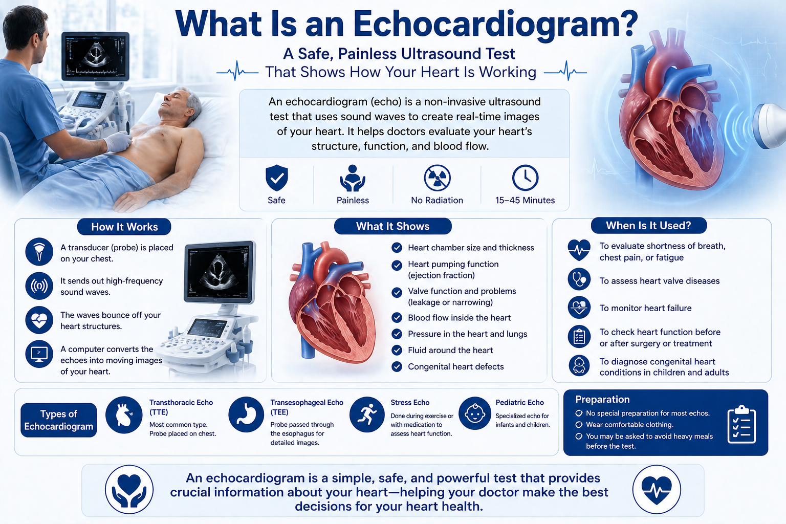
An echocardiogram — often called an “echo” — uses high-frequency sound waves (ultrasound) to produce real-time, moving images of your heart. Unlike an ECG (which records electrical signals), an echocardiogram shows the actual structure and movement of the heart: the walls, chambers, and valves — all in motion, in real time.
A standard echocardiogram takes 20–30 minutes, involves no radiation, no pain, and no preparation. A gel is applied to the chest, and a transducer (probe) is placed on the skin to capture images. An echocardiogram is completely safe for all ages including children and pregnant women.
What Does an Echocardiogram Show?
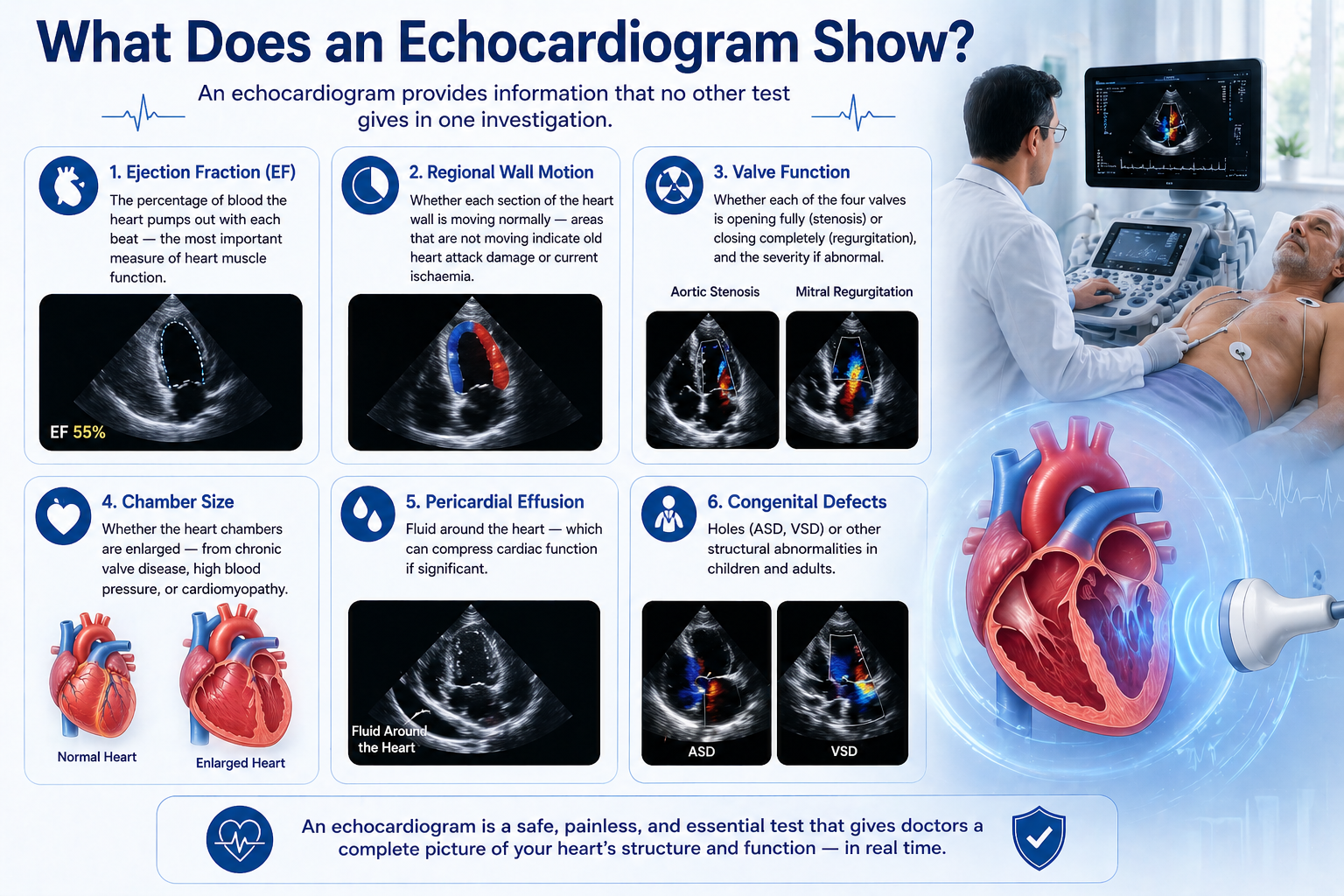
An echocardiogram provides information that no other test gives in one investigation:
- Ejection fraction (EF): The percentage of blood the heart pumps out with each beat — the most important measure of heart muscle function
- Regional wall motion: Whether each section of the heart wall is moving normally — areas that are not moving indicate old heart attack damage or current ischaemia
- Valve function: Whether each of the four valves is opening fully (stenosis) or closing completely (regurgitation), and the severity if abnormal
- Chamber size: Whether the heart chambers are enlarged — from chronic valve disease, high blood pressure, or cardiomyopathy
- Pericardial effusion: Fluid around the heart — which can compress cardiac function if significant
- Congenital defects: Holes (ASD, VSD) or other structural abnormalities in children and adults
How to Read Your Echocardiogram Report — Key Numbers Explained
Ejection Fraction (EF)
This is the most important number on an echocardiogram report. It measures the pumping power of the left ventricle.
- 55–70%: Normal
- 40–54%: Mildly reduced — warrants medication review and repeat echocardiogram
- 30–39%: Moderately reduced — significant heart failure; treatment and close monitoring required
- Below 30%: Severely reduced — requires specialist cardiac care and may change surgical timing and technique
Valve Grade (Severity)
Each valve abnormality is graded on the echocardiogram as mild, moderate, or severe. Mild disease is monitored. Moderate disease is reviewed every 6–12 months. Severe disease — particularly with symptoms or evidence of chamber enlargement — typically requires surgical discussion.
Wall Motion Score
Each segment of the left ventricular wall is graded from 1 (normal movement) to 4 (bulging paradoxically — indicating an aneurysm). Any score above 1 in multiple segments indicates past heart attack damage or ongoing ischaemia from blocked arteries.
Types of Echocardiogram
- Transthoracic echocardiogram (TTE): The standard echocardiogram — probe on the chest. Painless, no preparation, 20–30 minutes.
- Transoesophageal echocardiogram (TOE/TEE): A probe is passed into the oesophagus for much clearer images of certain structures — particularly the mitral valve and for detecting clots. Performed under sedation. Used before valve surgery and in suspected endocarditis.
- Stress echocardiogram: Echocardiogram performed during or immediately after exercise — reveals wall motion abnormalities that only appear when the heart is working hard. Used to detect blocked arteries when resting echocardiogram is normal.
- 3D echocardiogram: Three-dimensional reconstruction of the valve structure — increasingly used for surgical planning before mitral and tricuspid valve repair.
When Does an Echocardiogram Lead to Further Treatment?
An echocardiogram finding that typically leads to surgical consultation includes:
- Ejection fraction below 35% with symptoms of heart failure
- Severe aortic stenosis — particularly with symptoms
- Severe mitral regurgitation with increasing left ventricular dimensions
- Significant ASD or VSD with right heart enlargement
- Regional wall motion abnormality in a patient with symptoms — suggesting active ischaemia needing angiography
Dr. Ved Prakash reviews every echocardiogram in full before any cardiac surgical recommendation — not just the summary. Book a consultation for heart valve surgery in Delhi NCR or arrange an online cardiac consultation and share your echo report via WhatsApp.
Frequently Asked Questions — What Is an Echocardiogram
What is an echocardiogram and is it the same as an ECG?
No — an echocardiogram uses ultrasound to show the heart’s structure and movement. An ECG records only electrical signals. An echocardiogram gives vastly more information about valves, chamber size, and pumping function — it takes 20–30 minutes vs 5 minutes for an ECG.
What does a normal echocardiogram report show?
Normal echocardiogram findings: ejection fraction 55–70%, all four valves functioning normally, no wall motion abnormalities, normal chamber sizes, no fluid around the heart (no pericardial effusion).
What does ejection fraction mean on an echocardiogram?
Ejection fraction is the percentage of blood the left ventricle pumps out per beat. Normal is 55–70%. Below 40% indicates significant heart failure and influences decisions about surgery type and timing.
Is an echocardiogram safe?
Completely safe. An echocardiogram uses ultrasound, not radiation. No pain, no needles, no preparation for a standard study. Safe for children, pregnant women, and the elderly.
Dr. Ved Prakash | Director CTVS — Yatharth Super Speciality Hospitals, Greater Noida
📞 +91-9355255106 |
Book Appointment →