Author name: Atul Manori

Coronary Artery Disease
Uncategorized

Coronary Artery Disease — Stages, Symptoms and When Surgery Is Needed

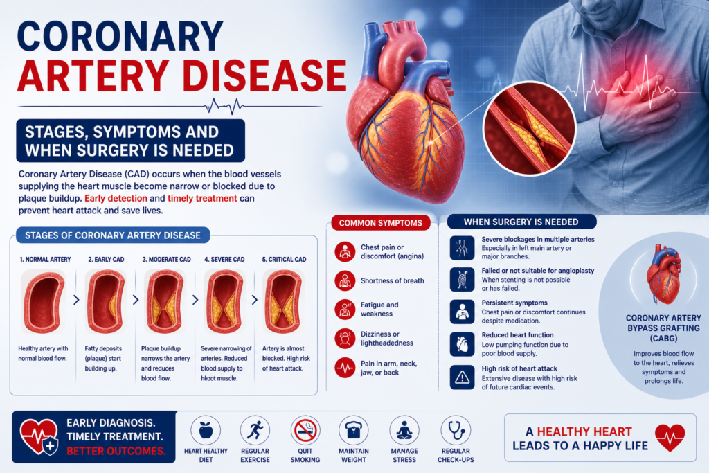
Coronary artery disease is not a single event — it is a decades-long process that progresses through identifiable stages, with each stage carrying different symptoms, different risks, and different treatment implications. Understanding where you or a family member sits in this progression is the most important step toward making the right treatment decision. Dr. Ved Prakash, Director of CTVS at Yatharth Super Speciality Hospitals, Greater Noida, explains coronary artery disease stages and symptoms, and when each stage crosses the threshold for surgical intervention.   What Is Coronary Artery Disease? Coronary artery disease (CAD) is the buildup of atherosclerotic plaque — a mixture of cholesterol, inflammatory cells, calcium, and fibrous tissue — inside the walls of the coronary arteries that supply the heart muscle with blood. As this plaque accumulates over years, it progressively narrows the artery lumen. When narrowing reaches approximately 70%, blood flow during exertion is insufficient and symptoms begin. When a plaque ruptures and a clot forms suddenly, the result is a heart attack. India has one of the highest burdens of coronary artery disease globally — affecting patients 10–15 years younger on average than in Western countries, and with a disproportionately high rate of multi-vessel disease at first presentation. Coronary Artery Disease Stages — The Full Progression Stage 1 — Subclinical Atherosclerosis (No Symptoms) Plaque begins accumulating in the coronary artery walls from as early as the 20s and 30s in high-risk individuals — those with diabetes, hypertension, high cholesterol, a strong family history, or heavy smoking history. At this stage, the narrowing is less than 50% and blood flow is not restricted. There are no symptoms whatsoever. This stage is detectable with CT coronary calcium scoring and CT coronary angiography — tests that show plaque burden before any symptoms develop. This is the stage where lifestyle modification and statin therapy are most effective at slowing or halting progression. Stage 2 — Stable Angina (Predictable Chest Pain on Exertion) When the coronary artery narrows beyond 70%, blood flow during physical exertion becomes insufficient for the heart muscle’s increased demand. The result is stable angina — chest pain or tightness that comes on predictably after a certain amount of exertion and resolves completely within 5–10 minutes of rest. Patients describe stable angina as: central chest tightness, pressure, heaviness, or aching — sometimes radiating to the left arm, jaw, or between the shoulder blades. The pain is consistent — the same exertion produces the same symptoms. It does not occur at rest. If you develop this symptom pattern, see a cardiologist immediately for an ECG, stress test, and likely coronary angiography. Diabetic patients and women frequently do not experience typical chest pain — they present with breathlessness, jaw pain, left arm pain, or unexplained fatigue as their angina equivalent. These atypical presentations lead to delayed diagnosis. Stage 3 — Unstable Angina / NSTEMI (Plaque Rupture) When atherosclerotic plaque becomes unstable — thin-capped, inflamed, and vulnerable — it can rupture. A ruptured plaque triggers immediate clot formation (thrombus) at the rupture site. If the clot partially blocks the artery, blood flow at rest is compromised. The result is: Unstable angina: Chest pain at rest, or with minimal exertion, or that is getting progressively more frequent and severe. A dangerous change from a previously stable pattern. NSTEMI (Non-ST Elevation Myocardial Infarction): Partial blockage with some heart muscle damage — troponin rises on blood tests but the ECG does not show the classic ST elevation of a full heart attack. Both unstable angina and NSTEMI require hospital admission and urgent coronary angiography — typically within 24–48 hours. This is not a situation where a patient should wait for a scheduled appointment. Stage 4 — STEMI (Complete Heart Attack) When the clot at the rupture site completely occludes the artery, blood supply to the downstream heart muscle stops entirely. This is a STEMI — ST-Elevation Myocardial Infarction — a full heart attack. Every minute of complete occlusion destroys heart muscle that will never recover. Time from symptom onset to opening the blocked artery is the single most important determinant of how much heart muscle is saved. STEMI symptoms: Sudden severe chest pain at rest — crushing, squeezing, or pressure sensation. May radiate to the left arm, jaw, or back. Often accompanied by sweating, nausea, breathlessness, and a feeling of impending doom. Can also present without chest pain in diabetics — only breathlessness and sweating. STEMI is a medical emergency. Call emergency services immediately. Do not drive yourself to hospital. How Many Arteries Are Blocked — And Why It Matters Coronary artery disease is classified by how many vessels are affected: Vessel Count Definition Typical Treatment Single vessel One artery with significant blockage Angioplasty usually appropriate Two vessel (double vessel) Two arteries significantly blocked Heart Team decision — depends on location and patient profile Three vessel (triple vessel) All three main arteries blocked Bypass surgery strongly preferred Left main disease Trunk artery before LAD and LCx Bypass surgery in most cases Risk Factors That Accelerate Coronary Artery Disease Diabetes — the single most aggressive accelerator of CAD in Indian patients. Doubles the risk and significantly worsens outcomes after heart attack. Hypertension — damages the arterial endothelium, accelerating plaque deposition Smoking — directly toxic to coronary endothelium; smokers develop CAD 10 years earlier than non-smokers High LDL cholesterol — primary driver of plaque accumulation; statin therapy is the cornerstone of prevention Family history — first-degree relative with heart disease before 55 (men) or 65 (women) doubles your risk Sedentary lifestyle and central obesity — particularly the android fat distribution pattern common in South Asians When Coronary Artery Disease Requires Surgery Not all coronary artery disease requires surgery. The threshold is determined by: Symptoms that are not adequately controlled with medication Anatomical findings on angiography that predict a survival benefit from surgery — triple vessel disease, left main disease, or high SYNTAX score Ejection fraction below 35% with multi-vessel disease — where complete revascularisation by surgery improves both symptoms and survival The decision between bypass

heart valve surgery cost in Delhi
Uncategorized

What Is TAVI? Heart Valve Replacement Without Open-Chest Surgery

TAVI — Transcatheter Aortic Valve Implantation — is a procedure that replaces the aortic heart valve through a catheter inserted through the leg artery, without opening the chest at all. It has transformed cardiac care for thousands of patients who were previously told they were too old or too unwell for heart surgery. Dr. Ved Prakash, Director of CTVS at Yatharth Super Speciality Hospitals, Greater Noida, explains the TAVI procedure, who qualifies, and how the decision between TAVI and open surgery is made.   What Is TAVI — The Basics TAVI (also called TAVR — Transcatheter Aortic Valve Replacement) is a catheter-based procedure that delivers a replacement aortic valve to the heart through an artery in the groin (femoral artery) — or sometimes through the chest wall (transapical) — without making a large chest incision or stopping the heart. The new biological tissue valve is compressed onto a small expandable frame and mounted on a catheter. Once positioned precisely inside the diseased native aortic valve using X-ray and echocardiographic guidance, the new valve is deployed — opening like a flower and immediately taking over the function of the diseased valve. The old, calcified valve leaflets are pushed aside. The procedure takes 1–2 hours. Most patients walk the next day and go home within 3–5 days. Why Was the TAVI Procedure Developed? Aortic stenosis — the narrowing of the aortic valve — is the most common serious valve disease in patients over 65. Untreated severe aortic stenosis carries an average survival of 1–3 years once symptoms appear. For decades, the only treatment was open-heart surgical valve replacement (SAVR) — which in elderly or medically frail patients carries significant risk. The TAVI procedure was developed specifically to offer these high-risk patients a life-saving valve replacement without the trauma of open surgery. First performed in 2002, TAVI has now been performed on over 1 million patients worldwide — with outcomes equivalent to surgery in high and intermediate-risk patients, and now expanding to low-risk patients as well. Who Is a Candidate for the TAVI Procedure? The TAVI procedure is recommended for patients with: Severe symptomatic aortic stenosis — confirmed on echocardiogram (valve area <1.0 cm², mean gradient >40 mmHg) AND symptoms of breathlessness, chest pain, or syncope High or intermediate surgical risk — assessed using the STS Score or EuroSCORE II (heart surgery risk calculators) Suitable aortic valve anatomy and femoral artery access — confirmed by CT angiography before the TAVI procedure The TAVI procedure is also being used increasingly in lower-risk patients — and as of the most recent ESC/ACC guidelines (2024), TAVI is a Class I recommendation across all risk groups for severe symptomatic aortic stenosis in patients over 75. TAVI vs Open Heart Surgery (SAVR) — How Is the Decision Made? This is where the Heart Team concept is essential — and where most TAVI articles fail patients by not explaining it. Every patient considered for aortic valve replacement at Yatharth Hospital is evaluated by a Heart Team — consisting of Dr. Ved Prakash (cardiac surgeon) + an interventional cardiologist + an anaesthetist — who review the echocardiogram, CT angiography, and clinical status together before recommending TAVI or surgery. Factor Favour TAVI Favour Open Surgery (SAVR) Age Over 75 years Under 65–70 years Surgical risk High or intermediate Low (young, fit patient) Lung function Poor (COPD, pulmonary fibrosis) Normal Aortic calcification Severe (avoids aortic clamping) Minimal Need for simultaneous procedures No — valve only Yes — coronary bypass + valve Valve durability need Less critical (older patient) Long term (younger patient) What Happens During the TAVI Procedure — Step by Step Anaesthesia: General anaesthesia or conscious sedation (local anaesthesia + sedation) — depending on the patient and the centre’s protocol Access: A small puncture in the femoral artery in the groin. No chest incision in transfemoral TAVI. Crossing the valve: A guidewire is advanced through the femoral artery, up the aorta, and across the diseased aortic valve — guided by X-ray Balloon valvuloplasty: A small balloon briefly opens the calcified valve to create space for the new valve Valve deployment: The replacement valve on its delivery system is positioned precisely at the level of the native aortic valve — then deployed. The valve immediately starts functioning. Confirmation: Echocardiography and angiography confirm the valve is working correctly, there is no significant leakage, and the coronary arteries remain open Access closure: The femoral artery puncture is closed with a vascular closure device — no surgical cut-down needed in most cases Life After the TAVI Procedure — What to Expect Hospital stay: 3–5 days in most patients — significantly shorter than the 7–10 days after open surgery Walking: The next day after the TAVI procedure in most cases Medications after TAVI: Aspirin lifelong (to prevent clotting on the new valve) + clopidogrel for 3–6 months. Warfarin is generally NOT required after TAVI unless you also have atrial fibrillation. Pacemaker: Approximately 10–15% of TAVI patients require a permanent pacemaker — the new valve can sometimes interfere with the heart’s electrical conduction system. This is discussed with every patient before the TAVI procedure. Echocardiogram follow-up: At 1 month, 1 year, and annually thereafter — to check valve function and detect any late complications TAVI valve lifespan: 10–15 years in most patients. If a TAVI valve wears out, a second TAVI (valve-in-valve) can often be performed. Frequently Asked Questions — What Is TAVI Procedure What is the TAVI procedure and is it major surgery? The TAVI procedure is a minimally invasive catheter-based aortic valve replacement — not open-heart surgery. No chest incision is made, the heart is not stopped, and no heart-lung machine is used. Most patients are discharged in 3–5 days and return to full activity within 2–4 weeks. Is TAVI safe for elderly patients? Yes — TAVI was specifically developed for high-risk and elderly patients who cannot safely undergo open surgery. In patients over 80 with severe aortic stenosis and multiple comorbidities, the TAVI procedure offers a survival benefit and quality-of-life improvement that would

peripheral artery disease symptoms
Uncategorized

Peripheral Artery Disease Symptoms — Why Leg Pain While Walking Is a Warning Sign

Peripheral artery disease symptoms are one of the most under-recognised warning signs in Indian medicine — and in diabetic patients, peripheral artery disease symptoms are particularly dangerous because they are often absent entirely until the disease is advanced. Dr. Ved Prakash, Director of CTVS and Vascular Surgery at Yatharth Super Speciality Hospitals, Greater Noida, explains peripheral artery disease symptoms, causes, risk groups, and when surgery or endovascular treatment is needed in Delhi NCR.   What Is Peripheral Artery Disease? Peripheral artery disease (PAD) is atherosclerosis of the arteries supplying the legs — the same process of plaque buildup that causes coronary artery disease in the heart, but affecting the vessels below the waist. As plaque builds up, the leg arteries narrow — reducing blood supply to the muscles and tissues of the leg. The result is the characteristic peripheral artery disease symptom of claudication: leg pain or cramping that comes on with walking and is relieved within 5 minutes of rest. Peripheral artery disease affects approximately 200 million people worldwide and is significantly underdiagnosed in India — particularly in patients with diabetes, where the classic peripheral artery disease symptoms may be absent due to accompanying nerve damage (neuropathy). Peripheral Artery Disease Symptoms — The Spectrum from Mild to Critical Stage 1 — Claudication (Intermittent Leg Pain on Walking) The hallmark peripheral artery disease symptom. Cramping, tightness, or aching pain in the calf, thigh, or buttock that begins predictably after a certain distance of walking — the claudication distance. The pain forces the patient to stop. After 5–10 minutes of rest, it completely resolves — and walking resumes. If you can walk 500 metres before the pain stops you, claudication is mild. If 100 metres stops you, claudication is severe. The location of the pain indicates the level of arterial disease: calf claudication means disease in the superficial femoral artery (mid-thigh level); thigh and buttock claudication indicates disease higher up, in the iliac arteries of the pelvis. Stage 2 — Reduced Claudication Distance (Disease Progression) As peripheral artery disease progresses, the claudication distance shortens. A patient who could walk 300 metres 2 years ago can now manage only 100 metres. This reflects worsening arterial narrowing — and is a signal that intervention should be considered before the next stage develops. Stage 3 — Rest Pain (Critical Ischaemia) When peripheral artery disease becomes severe enough, the foot no longer receives adequate blood even at rest. The patient experiences burning pain in the foot — classically worse at night, often requiring the foot to be hung over the edge of the bed (which uses gravity to increase blood flow). Rest pain is a medical emergency — limb loss occurs within weeks if not treated. Stage 4 — Tissue Loss (Gangrene or Ulceration) The most advanced peripheral artery disease symptom — wounds that do not heal, or areas of blackening (gangrene) on the toes or foot. In diabetic patients, a small foot wound that does not heal despite weeks of dressing is often the first presentation of severe underlying PAD. This stage requires urgent vascular surgery assessment — delay means amputation. Peripheral Artery Disease Symptoms in Diabetic Patients — The Silent Crisis Diabetic neuropathy damages the peripheral nerves — meaning diabetic patients with severe PAD may have no pain at all. They do not experience claudication, no rest pain, and they may not feel a wound developing on their foot. The first sign of advanced peripheral artery disease in a diabetic patient is often a non-healing ulcer or blackened toe — by which time the disease is already critical. All diabetic patients over 50 should have an annual ABI (Ankle-Brachial Index) measurement — a simple, painless test that compares blood pressure in the ankle to the arm to screen for peripheral artery disease silently. What Causes Peripheral Artery Disease? Peripheral artery disease is caused by atherosclerosis — the same risk factors as heart disease: Smoking: The single strongest modifiable risk factor for PAD — smokers have 3–5 times the peripheral artery disease risk of non-smokers. Stopping smoking slows PAD progression significantly. Diabetes: Doubles PAD risk and dramatically worsens outcomes — particularly for below-knee arterial disease Hypertension (high blood pressure) High cholesterol (hyperlipidaemia) Family history of vascular disease Obesity and sedentary lifestyle How Is Peripheral Artery Disease Diagnosed? ABI (Ankle-Brachial Index): Non-invasive, painless. A ratio below 0.9 confirms significant PAD. Below 0.5 indicates critical ischaemia. Duplex Doppler ultrasound: Maps the arterial anatomy and identifies the location and severity of narrowings CT angiography: Detailed 3D imaging of the arteries from abdomen to foot — essential for planning endovascular or surgical intervention Conventional angiography: Catheter-based — performed in the interventional suite when treatment (angioplasty or bypass) is planned immediately Peripheral Artery Disease Treatment in Delhi NCR Medical management: Antiplatelet drugs (aspirin or clopidogrel), statin therapy, blood pressure control, diabetes management, supervised exercise program (increases claudication distance by 50–100%), and strict smoking cessation Endovascular treatment (angioplasty/stenting): For suitable blockages — a balloon catheter opens the narrowed artery and a stent is placed to keep it open. Minimally invasive, no incision, 1–2 day hospital stay. Peripheral bypass surgery: For long or multiple blockages not suitable for endovascular treatment — a graft (either synthetic or vein graft from the leg) is used to bypass the blocked section. Effective and durable, with recovery of 4–6 weeks. For patients with critical ischaemia, urgent peripheral vascular disease treatment in Delhi NCR is available at Yatharth Super Speciality Hospitals, Greater Noida.

DVT deep vein thrombosis symptoms
Uncategorized

DVT deep vein thrombosis symptoms— Warning Signs and Treatment in India

EVLT laser varicose vein treatment is the most effective, least painful way to treat varicose veins in 2026 — a walk-in, walk-out laser procedure that closes the diseased vein from inside, with no incision, no stitches, and no general anaesthesia. Dr. Ved Prakash, Director of CTVS and Vascular Surgery at Yatharth Super Speciality Hospitals, Greater Noida, explains exactly how EVLT laser varicose vein treatment works, who qualifies, and what the recovery looks like.   What Is EVLT Laser Varicose Vein Treatment? EVLT stands for Endovenous Laser Treatment. In EVLT laser varicose vein treatment, a thin laser fibre is inserted directly into the incompetent saphenous vein (the main feeding vein of the varicosity) through a tiny needle puncture under ultrasound guidance. The laser fibre delivers controlled laser energy along the length of the vein as it is slowly withdrawn — causing the vein wall to collapse and seal permanently. Over the following 4–8 weeks, the sealed vein is absorbed by the body. Blood that previously pooled in the diseased vein is automatically rerouted through healthy deep veins — relieving symptoms and improving the appearance of the leg. How Is EVLT Laser Varicose Vein Treatment Performed? Step 1 — Duplex ultrasound mapping: Before EVLT, a duplex ultrasound scan maps the incompetent saphenous vein and its tributaries — identifying the extent of disease and planning where the laser fibre will be inserted Step 2 — Access: Under ultrasound guidance, a tiny needle puncture is made in the vein — usually just below the knee. No incision, no stitches. A thin sheath is inserted through which the laser fibre is passed up the length of the diseased vein. Step 3 — Tumescent anaesthesia: A dilute local anaesthetic solution (tumescent fluid) is injected around the vein using very fine needles. This serves two purposes — it numbs the area completely, and it acts as a heat barrier to protect the surrounding tissue from the laser energy. This is what makes EVLT laser varicose vein treatment painless. Step 4 — Laser activation: The laser is activated and the fibre is slowly pulled back along the vein — delivering energy uniformly to seal the entire length. This takes approximately 10–20 minutes. Step 5 — Compression: A compression bandage is applied immediately. The patient is asked to walk for 15–20 minutes before leaving the hospital — walking promotes healthy blood flow through the deep veins immediately after EVLT laser varicose vein treatment. Total procedure time including preparation: approximately 45–60 minutes. Performed under local anaesthesia. No overnight stay. Who Qualifies for EVLT Laser Varicose Vein Treatment? EVLT is suitable for: Symptomatic varicose veins from great or small saphenous vein incompetence CEAP Class C2–C6 — from symptomatic bulging veins to active venous ulcers Recurrent varicose veins after previous surgical stripping EVLT laser varicose vein treatment is not suitable for: Very large tortuous veins where the laser fibre cannot be advanced safely — surgical stripping may be needed Active DVT (deep vein thrombosis) in the treated leg Patients who cannot walk after the procedure (mobility is required for safe recovery) A duplex ultrasound assessment before EVLT laser varicose vein treatment confirms suitability in every case. Recovery After EVLT Laser Varicose Vein Treatment — Day by Day Day of procedure: Walk for 20–30 minutes immediately after. Go home. Mild tightness along the treated vein is normal. Days 1–3: Mild bruising and tightness along the treated vein — normal and expected. Walk 30 minutes daily. Wear compression stocking throughout the day. Avoid hot baths, saunas, and swimming. Days 4–7: Most patients return to desk work. Continue compression stockings during the day. Driving is permitted after Day 3 if the right leg was not treated — or when you can perform an emergency stop comfortably. Week 2: Bruising fades significantly. Tightness along the vein settles. Most daily activities fully resumed. Week 4–8: Duplex ultrasound check confirms the treated vein is fully closed and no re-canalisation has occurred. Visible varicose tributaries (smaller branches) may be treated with sclerotherapy at this visit if residual. Compression stockings: Worn for 2 weeks after EVLT laser varicose vein treatment — during the day only, removed at night. EVLT vs Surgical Stripping — Which Is Better? Feature EVLT Laser Treatment Surgical Stripping Anaesthesia Local anaesthesia General or spinal anaesthesia Incisions None — needle puncture only 2–3 small incisions Hospital stay Outpatient — same day Day procedure or overnight Return to work 1–2 days (desk work) 2–3 weeks Success rate at 5 years >90% vein closure >85% — similar Post-procedure pain Mild tightness only Moderate wound pain 5–7 days Frequently Asked Questions — EVLT Laser Varicose Vein Treatment Is EVLT laser varicose vein treatment painful? No — the tumescent anaesthesia numbs the entire vein before the laser is activated. Patients feel no pain during EVLT laser varicose vein treatment. Mild tightness along the treated vein is normal for 3–7 days after the procedure as the vein seals and is absorbed. How long does EVLT laser varicose vein treatment last? Vein closure rates at 5 years exceed 90% with EVLT laser varicose vein treatment. The treated vein is permanently sealed and absorbed by the body. New varicose veins may develop over subsequent years from other venous branches — particularly with risk factors like prolonged standing or pregnancy. Can both legs be treated with EVLT on the same day? Yes — bilateral EVLT laser varicose vein treatment is possible and commonly performed in a single session. Post-procedure walking is still required. Most patients tolerate bilateral EVLT well. What is the cost of EVLT laser varicose vein treatment in Delhi NCR? EVLT laser varicose vein treatment in Delhi NCR typically costs ₹40,000–₹80,000 per leg at an experienced vascular surgery centre. Insurance coverage is available when the procedure is performed for symptomatic varicose veins — check your policy for pre-authorisation requirements. Book an EVLT consultation for varicose veins in Delhi NCR at Yatharth Super Speciality Hospitals, Greater Noida. For more on varicose vein causes and progression, read our guide on varicose veins causes and

varicose veins causes symptoms
Uncategorized

Varicose Veins — Causes, Symptoms and Treatment Options in Delhi NCR

Varicose veins are not just a cosmetic problem — varicose veins causes include damaged vein valves that allow blood to pool in your legs, and without treatment, they can progress to skin ulcers, bleeding, and deep vein thrombosis. Dr. Ved Prakash, Director of CTVS and Vascular Surgery at Yatharth Super Speciality Hospitals, Greater Noida, explains varicose veins causes, symptoms, when they become medically serious, and what treatment options are available in Delhi NCR.   What Are Varicose Veins? Veins carry blood from the legs back to the heart — working against gravity. To prevent blood from flowing backwards, veins contain one-way valves that open when blood flows upward and close between heartbeats. When these valves weaken or are damaged, blood pools in the vein between beats — causing the vein to stretch, enlarge, and become twisted. The result is a varicose vein. Varicose veins are most common in the long saphenous vein system — the large superficial vein running from the foot to the groin along the inner leg. They appear as bulging, rope-like blue or purple veins visible under the skin, most commonly in the calf and thigh. Varicose Veins Causes — Why They Develop Understanding varicose veins causes helps identify who is most at risk. The primary mechanism is always weakened or incompetent vein valves — but several factors make this more likely: Primary Varicose Veins Causes Prolonged standing or sitting: The most significant occupational risk factor. Teachers, nurses, surgeons, security guards, factory workers, and retail staff who stand for 6+ hours daily have significantly higher rates of varicose vein causes. In sedentary workers, prolonged sitting compresses pelvic veins and impairs leg drainage. Genetics and family history: If one or both parents have varicose veins, your lifetime risk is 60–90%. Valve weakness tends to be inherited. Pregnancy: Multiple mechanisms — increased blood volume, hormonal relaxation of vein walls, and pressure on pelvic veins from the growing uterus. Varicose veins from pregnancy often improve after delivery but may persist and worsen with subsequent pregnancies. Obesity: Increased intra-abdominal pressure from excess weight impairs venous return from the legs. Age: Valve elasticity decreases naturally over time. Most patients with symptomatic varicose veins are over 40. Female sex: Women are twice as likely as men to develop varicose veins — due to hormonal effects on vein wall elasticity and the additional risk from pregnancy. Previous DVT (deep vein thrombosis): A past DVT can damage vein valves permanently — causing post-thrombotic varicose veins that are often more difficult to treat. Varicose Veins Symptoms — From Mild to Serious Varicose veins symptoms follow a progression described by the CEAP classification used by vascular surgeons worldwide: C1 — Spider veins: Small, thread-like red or purple veins visible near the skin surface. No symptoms — cosmetic only. C2 — Varicose veins: Bulging, rope-like veins. May or may not cause symptoms. C3 — Oedema (swelling): Ankle and leg swelling by evening that does not fully resolve overnight. Indicates significant venous hypertension. C4 — Skin changes: Skin discolouration (brown pigmentation), eczema, or hardening of the skin (lipodermatosclerosis) around the ankle. This indicates long-standing venous insufficiency. C5 — Healed venous ulcer: A healed ulcer — strongly associated with underlying varicose vein disease. C6 — Active venous ulcer: An open wound near the inner ankle that does not heal — a serious complication of untreated varicose veins. Requires urgent vascular surgery assessment. Symptoms at all stages include: heaviness and aching in the legs, worse by evening; itching over the veins; cramping at night; and visible swollen, twisted veins. The absence of visible veins does not rule out significant deep venous insufficiency — diagnosis requires a venous duplex ultrasound. When Do Varicose Veins Need Treatment? Not all varicose veins require treatment immediately. Treatment is recommended when: Symptoms significantly affect quality of life (aching, heaviness, cramps, swelling) Skin changes appear (C4) — indicating significant venous hypertension A venous ulcer is present or recurrent (C5–C6) Bleeding occurs from a superficial varicose vein Superficial thrombophlebitis (clot in a varicose vein causing redness and pain) recurs The patient wishes treatment for cosmetic reasons (any stage) Varicose Veins Treatment Options in Delhi NCR EVLT (Endovenous Laser Treatment): A laser fibre is inserted into the incompetent saphenous vein under local anaesthesia — the laser closes the vein from inside. No incision, walk-in walk-out procedure, return to work in 1–2 days. The preferred treatment for suitable varicose veins in Delhi NCR. RFA (Radiofrequency Ablation): Similar to EVLT but uses radiofrequency energy instead of laser. Equally effective, slightly less post-procedure bruising in some studies. Sclerotherapy: A chemical is injected directly into smaller varicose veins and spider veins — causing them to scar and close. Used for C1–C2 disease and residual small veins after EVLT. Surgical stripping: The saphenous vein is physically removed through small incisions under general anaesthesia. Effective but involves a longer recovery (2–3 weeks). Reserved for very large veins or when laser/RFA are not feasible. For a detailed look at EVLT, read our guide on EVLT laser treatment for varicose veins. Frequently Asked Questions — Varicose Veins Causes Symptoms What are the main varicose veins causes? The primary cause of varicose veins is weakened or incompetent one-way valves in the leg veins that allow blood to pool. The most significant risk factors are prolonged standing (occupational), family history, pregnancy, obesity, and increasing age. Women are twice as likely to develop varicose veins as men. Can varicose veins be dangerous? Yes — varicose veins can become medically dangerous when they cause skin ulcers (venous ulcers near the ankle), bleeding from a ruptured superficial vein, or when a clot forms (superficial thrombophlebitis). There is also an association between large varicose veins and increased DVT risk. Most cases are managed before these complications develop with appropriate treatment. Do varicose veins go away without treatment? No — varicose veins do not resolve on their own once established. Compression stockings reduce symptoms and slow progression, but they do not treat the underlying valve incompetence. The veins will gradually enlarge and symptoms

heart valve surgery cost in Delhi
Uncategorized

What Is Coronary Angiography? Procedure, Results and What Comes Next

Coronary angiography is the gold-standard test for identifying blockages in the heart arteries — the investigation that gives your cardiologist or cardiac surgeon the road map they need to plan the right treatment. If you have been referred for coronary angiography, or if you already have an angiography report and want to understand what it means, Dr. Ved Prakash, Director of CTVS at Yatharth Super Speciality Hospitals, Greater Noida, explains everything clearly.   What Is Coronary Angiography? Coronary angiography is a catheter-based procedure that injects a special dye (contrast agent) directly into the coronary arteries while X-ray images are taken continuously. The dye makes the inside of the arteries visible on X-ray — revealing exactly where blockages are, how severe they are, and how many arteries are affected. Coronary angiography is performed in a cardiac catheterisation laboratory (cath lab), takes approximately 30–45 minutes, and is done under local anaesthesia — you are awake throughout. The catheter is most commonly inserted through the radial artery at the wrist (radial access) — which causes much less discomfort and allows you to go home the same day or the following morning. Why Is Coronary Angiography Recommended? Your cardiologist recommends coronary angiography when: You have chest pain on exertion that suggests angina An ECG, stress test (TMT), or echocardiogram shows changes suggesting blocked arteries You are being evaluated before major non-cardiac surgery (e.g., valve surgery) and are over 50 or have risk factors You have had a heart attack and require urgent assessment of the blocked artery CT coronary angiography has shown suspected significant blockages that need catheter-based confirmation What Happens During Coronary Angiography — Step by Step Preparation: You fast for 4–6 hours before the procedure. A thin tube (IV line) is placed in your arm. The access site (wrist or groin) is cleaned and numbed with local anaesthetic. Catheter insertion: A thin flexible tube (catheter) is inserted into the artery and guided up to the heart — guided by X-ray imaging. You feel pressure but no pain. Dye injection: Contrast dye is injected into each coronary artery. You briefly feel a warm flush through the chest — this lasts 10–15 seconds and is completely normal. Images recorded: X-ray images (cine-angiograms) are taken from multiple angles as the dye flows through the arteries — revealing any narrowings or blockages. Catheter removal: The catheter is removed and pressure applied to the access site. With radial (wrist) access, a compression band is placed and you can sit up and eat within an hour. How to Read Your Coronary Angiography Report This is the section no competitor explains — yet it is what every patient needs to understand. Percentage Blockage (Stenosis) 0–49% stenosis: Mild narrowing — does not significantly restrict blood flow. No intervention needed. Managed with medication and lifestyle changes. 50–69% stenosis: Moderate narrowing — may or may not be causing symptoms. Sometimes assessed further with an FFR (Fractional Flow Reserve) pressure wire to determine if it is truly restricting flow. 70–90% stenosis: Significant blockage — restricts blood flow. Almost always treated with angioplasty (stent) or included in a bypass surgery plan. 90–99% stenosis: Critical blockage — very high risk of heart attack. Urgent treatment required. 100% (total occlusion): Complete blockage — the artery is fully closed. Treatment depends on how long it has been blocked and whether viable heart muscle is at risk. Which Arteries Are Named in the Report LAD (Left Anterior Descending): Supplies the front of the heart — the most important coronary artery, also called the “widow maker” LCx (Left Circumflex): Supplies the side and back of the heart RCA (Right Coronary Artery): Supplies the right ventricle and back of the left ventricle Left Main: The trunk from which the LAD and LCx branch — a critical vessel; significant blockage here is a cardiac emergency What Happens After Coronary Angiography If a Blockage Is Found? Single, simple blockage in a non-diabetic patient: May be treated with angioplasty (stenting) immediately in the same session or scheduled shortly after Multiple blockages or complex anatomy: The coronary angiography images are reviewed by a Heart Team — cardiologist and cardiac surgeon — to decide between angioplasty and bypass surgery based on the SYNTAX score Left main disease: A cardiac surgery review is mandatory — bypass surgery is usually preferred No significant blockages: The angiography rules out coronary artery disease — your chest symptoms need evaluation for other causes If you have had coronary angiography and have been told surgery is needed, a cardiac second opinion in Delhi NCR from Dr. Ved Prakash gives you a complete, independent review of your angiogram images and a clear treatment recommendation. Share your angiography CD and report via WhatsApp. Frequently Asked Questions — What Is Coronary Angiography What is coronary angiography and is it dangerous? Coronary angiography is a catheter-based diagnostic procedure using dye and X-ray to reveal coronary artery blockages. It is extremely safe — the risk of serious complication is less than 0.1% in elective cases. It is performed under local anaesthesia and takes 30–45 minutes. What does 70% blockage on angiography mean? A 70% stenosis means the artery is narrowed to 30% of its normal diameter — significantly restricting blood flow. Most cardiologists and cardiac surgeons recommend treatment (angioplasty or bypass, depending on location) for blockages of 70% or more in major coronary arteries. Is coronary angiography painful? Not painful. The access site is numbed with local anaesthetic. Patients feel brief pressure when the catheter is moved, and a warm flush when the dye is injected — both normal and temporary. Most patients find coronary angiography significantly less uncomfortable than anticipated. What happens after coronary angiography if a blockage is found? A single simple blockage may be stented in the same session. Multiple or complex blockages are reviewed by a Heart Team who recommend angioplasty or bypass surgery based on the SYNTAX score and patient factors. Dr. Ved Prakash | Director CTVS — Yatharth Super Speciality Hospitals, Greater Noida 📞 +91-9355255106  | Bypass Surgery Consultation

Uncategorized

What Is an Echocardiogram? What It Shows and Why Your Doctor Ordered It

An echocardiogram is an ultrasound scan of your heart — the single most important investigation in cardiac medicine, and the test that Dr. Ved Prakash reviews before making any treatment decision. If your doctor has ordered an echocardiogram, this article explains what it is, how to read the key results, and what the findings may mean for your treatment at Yatharth Super Speciality Hospitals, Greater Noida.   What Is an Echocardiogram — The Basics An echocardiogram — often called an “echo” — uses high-frequency sound waves (ultrasound) to produce real-time, moving images of your heart. Unlike an ECG (which records electrical signals), an echocardiogram shows the actual structure and movement of the heart: the walls, chambers, and valves — all in motion, in real time. A standard echocardiogram takes 20–30 minutes, involves no radiation, no pain, and no preparation. A gel is applied to the chest, and a transducer (probe) is placed on the skin to capture images. An echocardiogram is completely safe for all ages including children and pregnant women. What Does an Echocardiogram Show? An echocardiogram provides information that no other test gives in one investigation: Ejection fraction (EF): The percentage of blood the heart pumps out with each beat — the most important measure of heart muscle function Regional wall motion: Whether each section of the heart wall is moving normally — areas that are not moving indicate old heart attack damage or current ischaemia Valve function: Whether each of the four valves is opening fully (stenosis) or closing completely (regurgitation), and the severity if abnormal Chamber size: Whether the heart chambers are enlarged — from chronic valve disease, high blood pressure, or cardiomyopathy Pericardial effusion: Fluid around the heart — which can compress cardiac function if significant Congenital defects: Holes (ASD, VSD) or other structural abnormalities in children and adults How to Read Your Echocardiogram Report — Key Numbers Explained Ejection Fraction (EF) This is the most important number on an echocardiogram report. It measures the pumping power of the left ventricle. 55–70%: Normal 40–54%: Mildly reduced — warrants medication review and repeat echocardiogram 30–39%: Moderately reduced — significant heart failure; treatment and close monitoring required Below 30%: Severely reduced — requires specialist cardiac care and may change surgical timing and technique Valve Grade (Severity) Each valve abnormality is graded on the echocardiogram as mild, moderate, or severe. Mild disease is monitored. Moderate disease is reviewed every 6–12 months. Severe disease — particularly with symptoms or evidence of chamber enlargement — typically requires surgical discussion. Wall Motion Score Each segment of the left ventricular wall is graded from 1 (normal movement) to 4 (bulging paradoxically — indicating an aneurysm). Any score above 1 in multiple segments indicates past heart attack damage or ongoing ischaemia from blocked arteries. Types of Echocardiogram Transthoracic echocardiogram (TTE): The standard echocardiogram — probe on the chest. Painless, no preparation, 20–30 minutes. Transoesophageal echocardiogram (TOE/TEE): A probe is passed into the oesophagus for much clearer images of certain structures — particularly the mitral valve and for detecting clots. Performed under sedation. Used before valve surgery and in suspected endocarditis. Stress echocardiogram: Echocardiogram performed during or immediately after exercise — reveals wall motion abnormalities that only appear when the heart is working hard. Used to detect blocked arteries when resting echocardiogram is normal. 3D echocardiogram: Three-dimensional reconstruction of the valve structure — increasingly used for surgical planning before mitral and tricuspid valve repair. When Does an Echocardiogram Lead to Further Treatment? An echocardiogram finding that typically leads to surgical consultation includes: Ejection fraction below 35% with symptoms of heart failure Severe aortic stenosis — particularly with symptoms Severe mitral regurgitation with increasing left ventricular dimensions Significant ASD or VSD with right heart enlargement Regional wall motion abnormality in a patient with symptoms — suggesting active ischaemia needing angiography Dr. Ved Prakash reviews every echocardiogram in full before any cardiac surgical recommendation — not just the summary. Book a consultation for heart valve surgery in Delhi NCR or arrange an online cardiac consultation and share your echo report via WhatsApp. Frequently Asked Questions — What Is an Echocardiogram What is an echocardiogram and is it the same as an ECG? No — an echocardiogram uses ultrasound to show the heart’s structure and movement. An ECG records only electrical signals. An echocardiogram gives vastly more information about valves, chamber size, and pumping function — it takes 20–30 minutes vs 5 minutes for an ECG. What does a normal echocardiogram report show? Normal echocardiogram findings: ejection fraction 55–70%, all four valves functioning normally, no wall motion abnormalities, normal chamber sizes, no fluid around the heart (no pericardial effusion). What does ejection fraction mean on an echocardiogram? Ejection fraction is the percentage of blood the left ventricle pumps out per beat. Normal is 55–70%. Below 40% indicates significant heart failure and influences decisions about surgery type and timing. Is an echocardiogram safe? Completely safe. An echocardiogram uses ultrasound, not radiation. No pain, no needles, no preparation for a standard study. Safe for children, pregnant women, and the elderly. Dr. Ved Prakash | Director CTVS — Yatharth Super Speciality Hospitals, Greater Noida 📞 +91-9355255106  | Book Appointment →

Questions to Ask Your Cardiac Surgeon
Uncategorized

On-Pump vs Off-Pump Bypass Surgery — Which Is Better for You?

On-pump vs off-pump bypass surgery is one of the most specific questions patients ask before CABG — and it is a genuinely important one, because the right technique depends directly on your individual health profile. Dr. Ved Prakash, Director of CTVS at Yatharth Super Speciality Hospitals, Greater Noida, has extensive experience with both techniques and explains how the on-pump vs off-pump bypass surgery decision is made — and what it means for your recovery.   What Is On-Pump Bypass Surgery? In on-pump bypass surgery, the heart is temporarily stopped using a special medication (cardioplegia solution), and a heart-lung machine (cardiopulmonary bypass machine) takes over the job of circulating and oxygenating the blood while the surgeon operates. Because the heart is completely still, the surgeon can work with great precision in placing the bypass grafts. On-pump bypass surgery is the standard, time-tested technique that has been performed since the 1960s. It remains the default approach for most complex CABG operations worldwide — and it delivers excellent, consistent outcomes. What Is Off-Pump Bypass Surgery (OPCAB)? In off-pump bypass surgery — also called OPCAB or beating-heart bypass — the bypass grafts are attached while the heart continues to beat. No heart-lung machine is used. The surgeon uses mechanical stabilisers to gently immobilise a small area around the target coronary artery while the rest of the heart beats normally. Off-pump bypass surgery is technically more demanding — it requires greater surgical skill and experience than the standard on-pump technique. But in the right patients and the right hands, it offers meaningful advantages. On-Pump vs Off-Pump Bypass Surgery — Key Differences Feature On-Pump CABG Off-Pump CABG (OPCAB) Heart-lung machine used? Yes No Heart stopped during surgery? Yes No — heart continues to beat Surgical complexity Standard Higher — requires specialised technique Kidney complication risk Slightly higher Reduced in pre-existing kidney disease Stroke risk Slightly higher in aortic calcification Potentially lower in high-risk patients Blood transfusion need Slightly higher Often lower Graft completeness Easier to achieve all targets Some posterior targets technically harder Overall outcome Excellent Equivalent in experienced hands Who Benefits Most From Off-Pump Bypass Surgery? On-pump vs off-pump bypass surgery — the off-pump technique has specific advantages for certain patient groups: Patients with chronic kidney disease (CKD): The heart-lung machine can reduce kidney blood flow during surgery — off-pump bypass avoids this and is strongly preferred when baseline kidney function is already reduced Patients with a heavily calcified aorta: Clamping the aorta for the heart-lung machine in a calcified vessel can dislodge calcium fragments and cause stroke — off-pump surgery avoids aortic clamping in selected techniques Patients with significant lung disease (COPD): The heart-lung machine can worsen lung inflammation; off-pump bypass reduces this risk Elderly patients: Reduced exposure to the inflammatory effects of cardiopulmonary bypass may benefit older patients Jehovah’s Witnesses or patients refusing blood transfusion: Off-pump bypass surgery typically results in less blood loss Who Is Better Suited to On-Pump Bypass Surgery? In the on-pump vs off-pump bypass surgery comparison, on-pump is preferred when: The coronary arteries to be bypassed are in technically difficult positions (particularly posterior vessels on the back of the heart) that are harder to stabilise in a beating heart The heart is already significantly enlarged or weak — making stabilisation for off-pump technically challenging The operation needs to be combined with another procedure simultaneously (e.g., valve repair or aortic surgery) The surgeon’s experience is primarily with the on-pump technique — surgeon experience matters more than technique choice   The Most Important Factor — Surgeon Experience The on-pump vs off-pump bypass surgery debate in the medical literature largely resolves to one conclusion: in experienced hands, both techniques deliver equivalent long-term graft patency and mortality rates. The difference is in short-term kidney and neurological complication rates — which benefit specific patient subgroups with off-pump surgery. What this means practically: the technique that is safer for you is the one your surgeon has the most experience with, applied to your specific anatomy and health profile. Dr. Ved Prakash discusses this choice openly with every patient before surgery. Frequently Asked Questions — On-Pump vs Off-Pump Bypass Surgery What is the difference between on-pump and off-pump bypass surgery? In on-pump bypass surgery, a heart-lung machine temporarily takes over the heart’s function while the surgeon operates on a still heart. In off-pump bypass surgery, the grafts are attached while the heart continues to beat — no heart-lung machine is used. Both produce excellent outcomes in the right patient. Is off-pump bypass surgery safer than on-pump? Off-pump bypass surgery reduces kidney complication risk and may lower stroke risk in high-risk patients. However, it is technically more demanding and requires greater surgical experience. In experienced hands, both techniques deliver equivalent overall outcomes for most patients. Who is off-pump bypass surgery best for? Patients with pre-existing kidney disease, heavily calcified aortas, significant lung disease, or those at elevated risk of neurological complications from the heart-lung machine benefit most from off-pump bypass surgery. Can all blockages be bypassed in off-pump surgery? Most coronary artery blockages can be bypassed off-pump by an experienced surgeon. However, certain anatomical locations — particularly vessels on the back of the heart — are more challenging. If complete revascularisation cannot be achieved off-pump, conversion to on-pump surgery is performed without any compromise to safety. For more on bypass surgery in general, read our complete guide on bypass surgery vs angioplasty — or book a consultation for bypass surgery in Delhi NCR. Dr. Ved Prakash | Director CTVS — Yatharth Super Speciality Hospitals, Greater Noida 📞 +91-9355255106  | Book Appointment →

Bypass Surgery vs Angioplasty
Uncategorized

Bypass Surgery vs Angioplasty — How Is the Decision Made?

The bypass surgery vs angioplasty decision is the most important — and most frequently misunderstood — choice in cardiac treatment. Both procedures treat blocked coronary arteries. Both save lives. But choosing the wrong one for your anatomy can mean going back for another procedure in 5 years — or worse. Dr. Ved Prakash, Director of CTVS at Yatharth Super Speciality Hospitals, Greater Noida, explains exactly how bypass surgery vs angioplasty is evaluated — and what determines the right answer for each patient.   Bypass Surgery vs Angioplasty — What Each Procedure Does Before comparing bypass surgery vs angioplasty, the basic mechanism of each must be clear. Angioplasty (PCI / stenting): A thin catheter is inserted through the wrist or groin artery and guided to the blockage. A balloon inflates to widen the artery, and a metal mesh tube (stent) is left in place to keep it open. No chest incision. Recovery in 1–3 days. Best for: a single blockage or a simple two-vessel disease in a non-diabetic patient. Bypass surgery (CABG): A healthy blood vessel is taken from the chest wall or leg and sewn in to bypass the blocked section entirely — creating a permanent new route for blood. Requires a chest incision and 7–10 days in hospital. Best for: multiple blockages, left main disease, diabetes, or complex anatomy that stents cannot address durably. The Clinical Criteria Used to Choose Between Bypass Surgery vs Angioplasty The bypass surgery vs angioplasty decision is not made on a single factor. It requires reviewing the angiogram against a structured set of criteria. At Yatharth Hospital, every complex case is reviewed by both Dr. Ved Prakash (cardiac surgeon) and an interventional cardiologist before a recommendation is made — this is called the Heart Team approach, and it is the international standard. 1. Number of Blocked Arteries Single-vessel disease: Angioplasty is usually appropriate if the lesion is technically suitable Two-vessel disease: May be treated with either — depends on location and patient factors Three-vessel disease (triple vessel disease): Bypass surgery vs angioplasty — bypass surgery is strongly preferred. Multiple stents in all three arteries have higher restenosis rates and do not match the durability of a triple bypass 2. Left Main Coronary Artery Disease The left main artery supplies 70% of the heart muscle. A significant blockage here is a surgical emergency — or at minimum, a case where bypass surgery vs angioplasty must be carefully weighed by a Heart Team. Current guidelines recommend bypass surgery for most left main disease, particularly when combined with other vessel disease. 3. Diabetes This is the single most important factor that tips the bypass surgery vs angioplasty decision toward bypass for multi-vessel disease. The landmark FREEDOM trial showed that diabetic patients with multi-vessel disease who received bypass surgery had significantly lower mortality and heart attack rates at 5 years compared to angioplasty. Diabetic vessels tend to re-block stents at higher rates — making the more durable bypass graft the better long-term choice. 4. The SYNTAX Score The SYNTAX score is an angiographic scoring system that quantifies the complexity of coronary artery blockages. A higher score means more complex disease. In general: Low SYNTAX score (0–22): Either bypass surgery or angioplasty — angioplasty outcomes are equivalent Intermediate SYNTAX score (23–32): Individualised decision — Heart Team required High SYNTAX score (33+): Bypass surgery is strongly preferred — angioplasty outcomes are significantly inferior 5. Heart Muscle Function (Ejection Fraction) When the heart’s pumping function is significantly reduced (ejection fraction below 35%), bypass surgery has been shown to improve survival more reliably than angioplasty — because restoring complete blood supply to the struggling heart muscle requires grafting all diseased vessels, which multiple stents rarely achieve as completely. 6. Age and Surgical Fitness For very elderly patients (over 80) or those with severe lung disease, kidney failure, or other conditions that make surgery high-risk — angioplasty may be preferred even when bypass surgery would normally be the better anatomical choice. The bypass surgery vs angioplasty decision always accounts for the patient’s overall health, not just the angiogram. Bypass Surgery vs Angioplasty — Side-by-Side Comparison Factor Bypass Surgery (CABG) Angioplasty (PCI) Recovery time 6–12 weeks 1–3 days Best for diabetes + multi-vessel Yes — strongly preferred Higher restenosis risk Left main disease Preferred in most cases Only in selected anatomy Graft / stent durability LIMA graft: 15–20+ years Drug-eluting stent: 10–15 years Three-vessel disease Strongly preferred High SYNTAX score → inferior outcomes Blood thinners needed Aspirin only (lifelong) Aspirin + Clopidogrel (12 months minimum) Can be repeated if it fails Yes, but higher risk redo Yes, repeat angioplasty or bypass When Bypass Surgery vs Angioplasty Is Not Clear-Cut — Get a Second Opinion If your cardiologist has recommended angioplasty for multi-vessel disease and you are diabetic — or if you have been offered bypass surgery and want to understand whether angioplasty is viable — a cardiac second opinion in Delhi NCR from a CTVS surgeon who reviews your angiogram independently is the most important step before making a decision. Share your angiogram CD and report via WhatsApp with Dr. Ved Prakash for a pre-assessment. Frequently Asked Questions — Bypass Surgery vs Angioplasty Is bypass surgery vs angioplasty always a surgeon’s decision? No — the decision should be made jointly by an interventional cardiologist and a cardiac surgeon reviewing the angiogram together. A Heart Team approach using the SYNTAX score is the international standard. Any patient offered one option without this joint review should ask why. Which is better — bypass surgery or angioplasty? For single, simple blockages in otherwise healthy patients — angioplasty is usually sufficient. For three-vessel disease, left main disease, or diabetic patients with multiple blockages — bypass surgery gives more durable long-term results. There is no universal winner; the anatomy of the blockages decides. Can bypass surgery be done after failed angioplasty? Yes — bypass surgery is commonly performed after a stent fails (restenosis) or when multiple stents have been placed and further angioplasty is no longer feasible. It

Uncategorized

Life After Bypass Surgery — What to Expect Week by Week

Life after bypass surgery is significantly better for most patients than life before it — less chest pain, better breathing, more energy, and a reduced risk of heart attack. But the weeks between leaving the operating theatre and reaching full recovery require patience, the right information, and realistic expectations. Dr. Ved Prakash, Director of CTVS at Yatharth Super Speciality Hospitals, Greater Noida, has guided hundreds of patients through bypass surgery recovery. This is what actually happens, week by week.   Before We Begin — What Most Patients Feel in the First Days Immediately after bypass surgery, most patients are surprised by three things. First, how alert they feel once the anaesthesia wears off. Second, that the breathing tube is usually removed within 6–8 hours — far sooner than they expected. Third, that they are asked to sit up and take a few steps as early as Day 2. Recovery from bypass surgery is faster than most patients imagine — because the goal throughout is to get the heart and body moving again as soon as safely possible. Week-by-Week Bypass Surgery Recovery Timeline Days 1–2: Cardiac ICU You wake up in the cardiac ICU with monitoring lines and a breathing tube — both are removed within hours once you are stable Pain is managed continuously with IV medications — the nurses ask about your pain level regularly A physiotherapist visits on Day 1 to guide you through breathing exercises — these are critical for preventing lung complications You will be sitting up in a chair by Day 2 in most cases Chest drain tubes are usually removed on Day 2 Days 3–7: Hospital Ward You are moved from ICU to the general cardiac ward once stable Short walks in the corridor begin — typically 50–100 metres initially, increasing each day Eating and drinking normally resumes The chest wound is checked and dressed daily Medications are reviewed and finalised before discharge — aspirin, statins, beta-blocker are standard Most patients are discharged on Day 7–10 Weeks 2–4: Home Recovery The sternum (breastbone) is healing — it takes 6–8 weeks to fully unite. During this time, do not push yourself up from chairs using your arms, do not lift anything heavier than a glass of water, and do not drive Walk daily — start with 5–10 minutes and increase by 2–3 minutes each day as tolerated Shower is permitted once wounds are dry and sealed — usually by Day 10–12 Sleep is often interrupted in the first 2–3 weeks — this is normal and temporary Mild depression or emotional low is common in the first 2–3 weeks — it resolves as energy and confidence return First outpatient follow-up is at 2 weeks for wound check and blood tests Weeks 5–6: Healing Milestone The sternum is now mostly healed — gentle arm use and light daily activities resume Walking 20–30 minutes daily is the goal by Week 6 Most patients notice a real improvement in how they feel — chest pain is gone, breathing is easier, energy is returning You may return to light desk work from home if energy allows Driving is permitted at 6 weeks if the sternum feels stable and you can perform an emergency stop without hesitation Weeks 7–12: Return to Normal Life Most patients return to their regular routine — including work — between 8 and 12 weeks Cardiac rehabilitation (supervised exercise program) typically begins at Week 6–8 — strongly recommended for all bypass surgery patients Sexual activity can usually resume at Week 6–8 when sternum is fully healed Air travel is generally safe at 6–8 weeks with medical clearance Lifting heavier objects — up to 5 kg — is usually permitted by Week 8–10 What You Should Not Do After Bypass Surgery Activity When It Is Safe Driving 6 weeks minimum Lifting over 5 kg 8–10 weeks Return to desk work 6–8 weeks Return to physical work 12 weeks minimum Swimming 12 weeks (chest wound must be fully healed) Air travel 6–8 weeks with medical clearance Medications After Bypass Surgery — What and Why Most bypass surgery patients are discharged on the following medications — do not stop any of them without consulting your doctor: Aspirin: Keeps the bypass grafts open by preventing clotting. Lifelong. Statin (e.g. Atorvastatin): Controls cholesterol and protects the arteries. Lifelong. Beta-blocker (e.g. Metoprolol): Controls heart rate and reduces cardiac workload. Usually for at least 12 months, often longer. ACE inhibitor (e.g. Ramipril): Protects heart muscle function. Often long-term especially if ejection fraction was low. Warning Signs After Bypass Surgery — When to Seek Help Immediately High fever (above 101°F / 38.5°C) — may indicate wound infection Redness, swelling, or discharge from the chest wound or leg wound Chest pain — new or different from before surgery Significant breathlessness at rest Leg swelling or calf pain — possible DVT Sudden weakness or slurred speech — seek emergency care immediately Frequently Asked Questions — Life After Bypass Surgery How long does it take to fully recover from bypass surgery? Most patients feel significantly better by 6 weeks and return to full normal life by 8–12 weeks. The sternum takes 6–8 weeks to fully heal — during this period physical restrictions apply. After 12 weeks, there are generally no long-term restrictions on activity. Will I feel better after bypass surgery than before? For the vast majority of patients — yes, dramatically. The relief of restored blood flow to the heart eliminates angina completely in most cases. Patients frequently report being able to walk, climb stairs, and exercise without chest pain or breathlessness for the first time in years. Is depression common after bypass surgery? Yes — a significant proportion of bypass surgery patients experience mild depression or emotional low in the first 2–4 weeks after surgery. This is partly hormonal (the body’s response to major surgery) and partly psychological. It almost always resolves as physical recovery progresses. Talk to your cardiac team if it persists beyond 4–6 weeks. How long do bypass grafts last? The LIMA

Scroll to Top